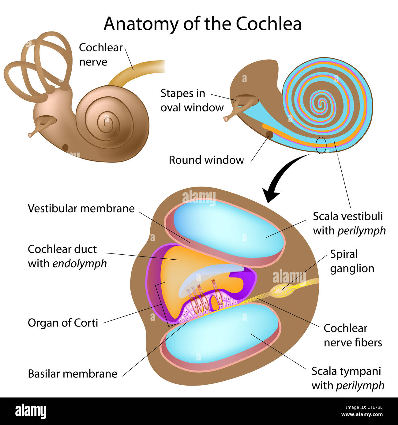
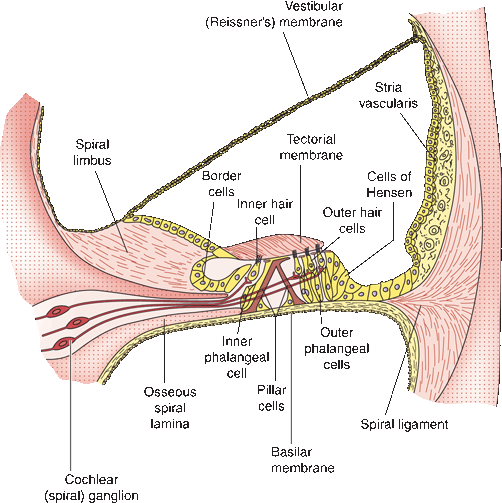
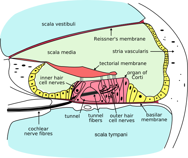
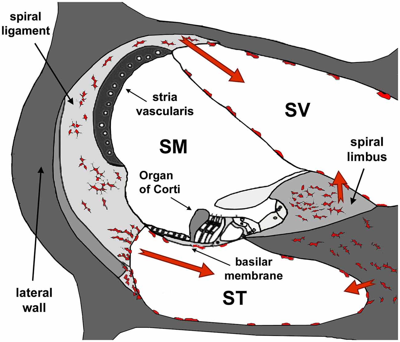
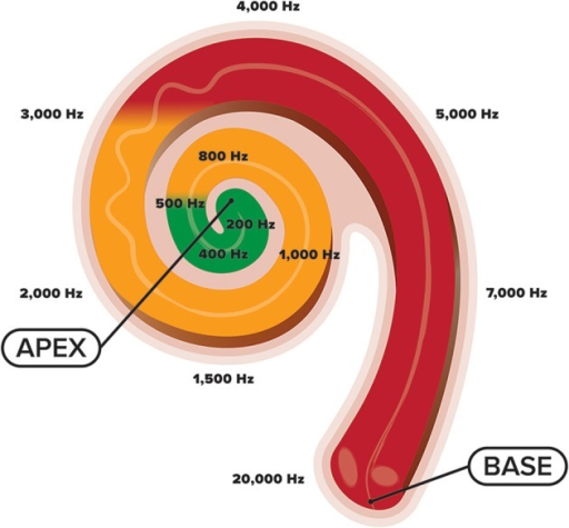
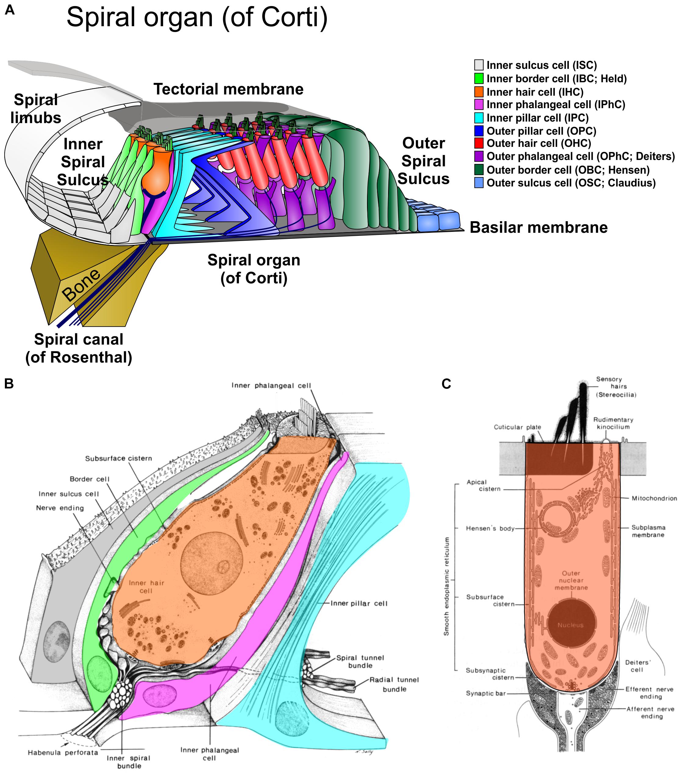
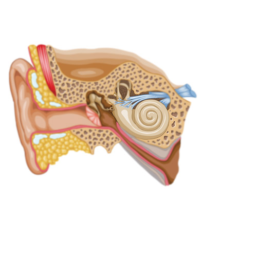
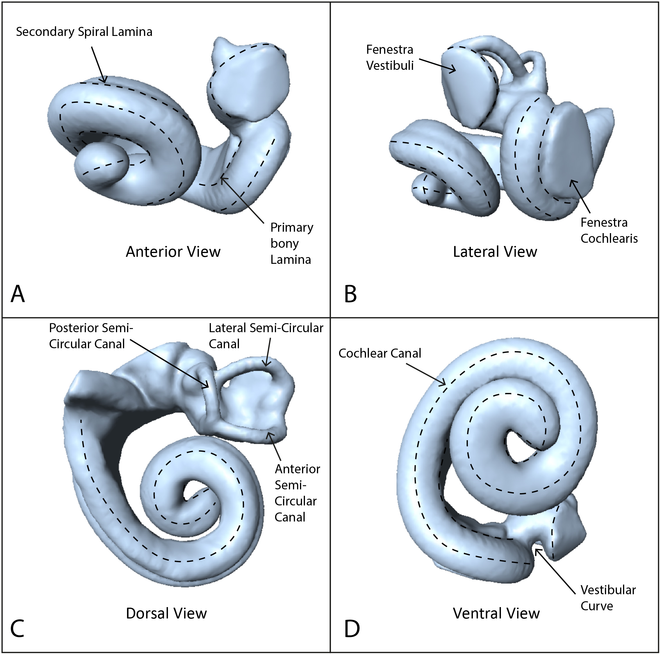
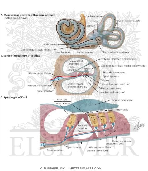
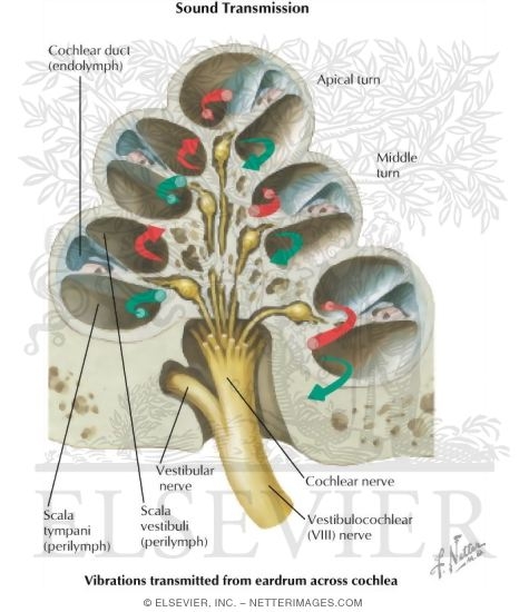
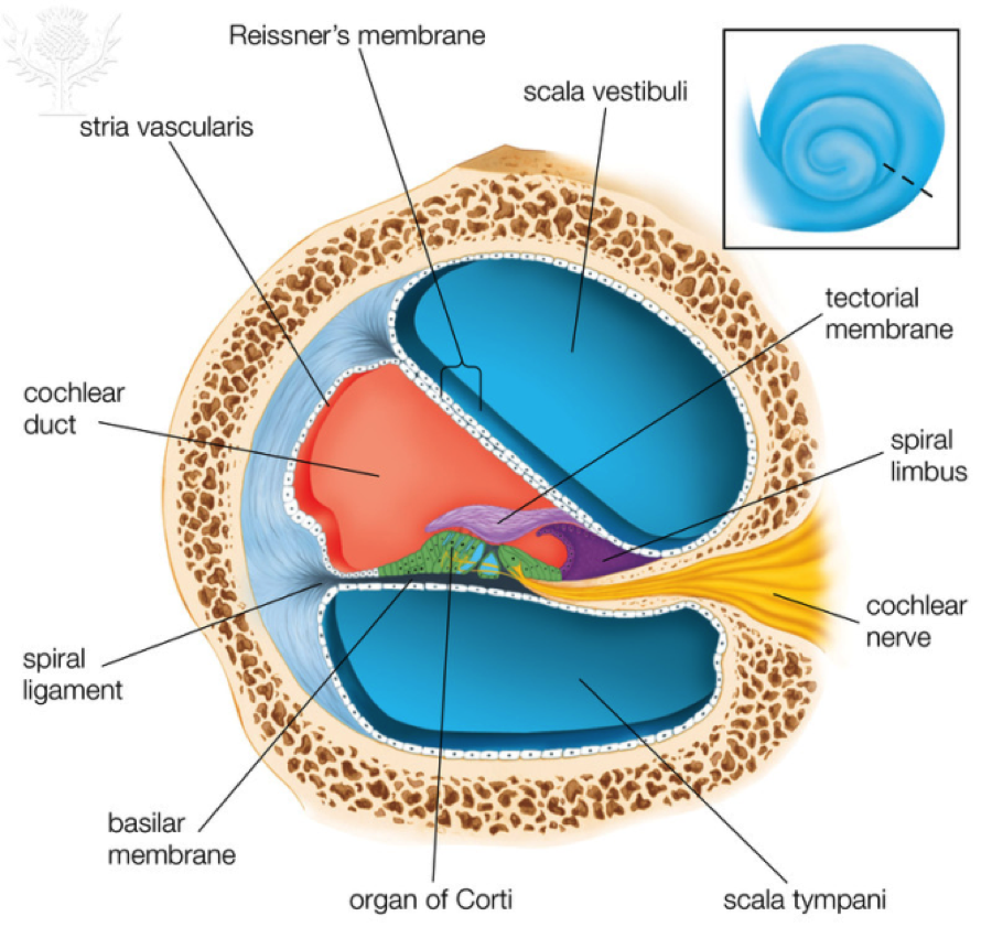
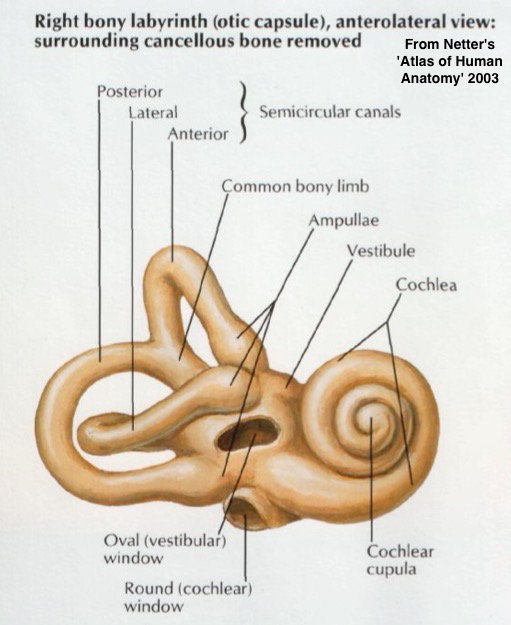
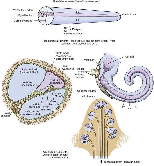
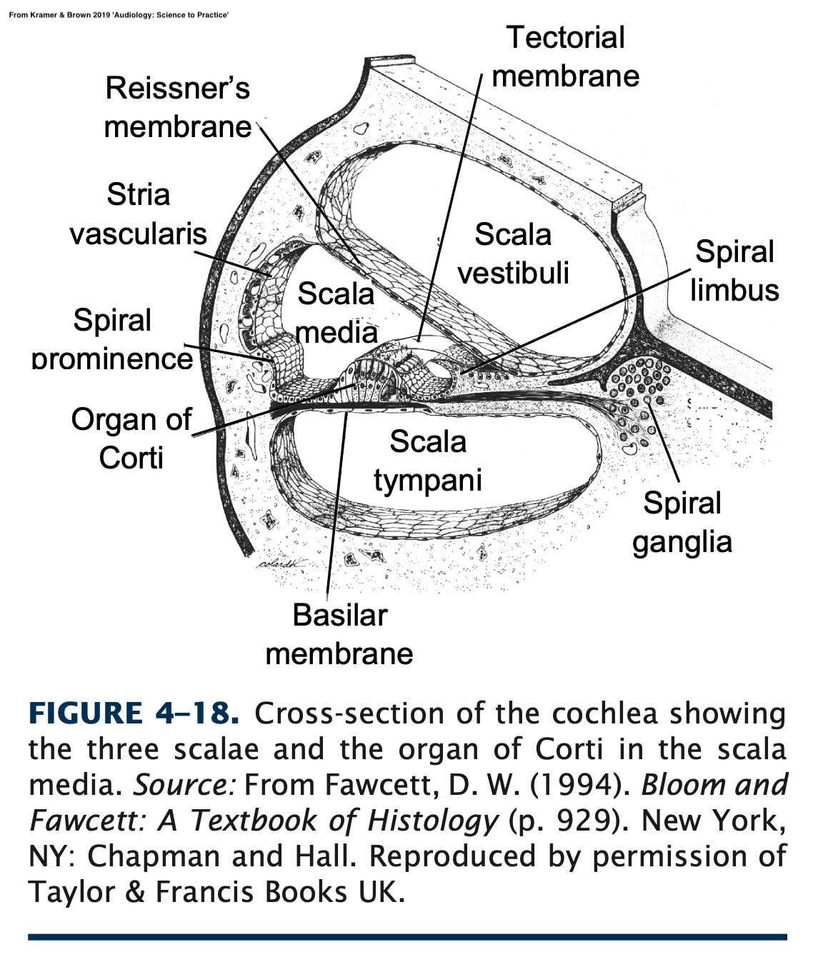
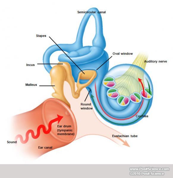
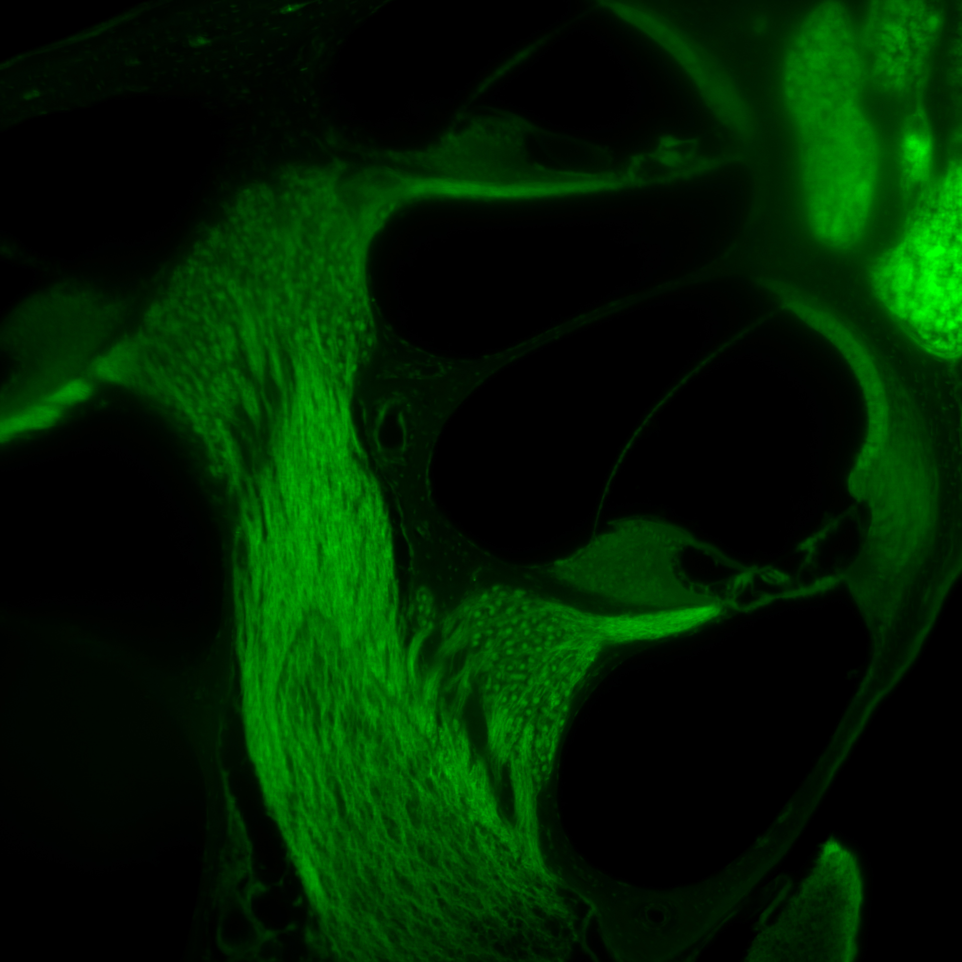
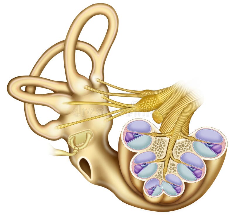
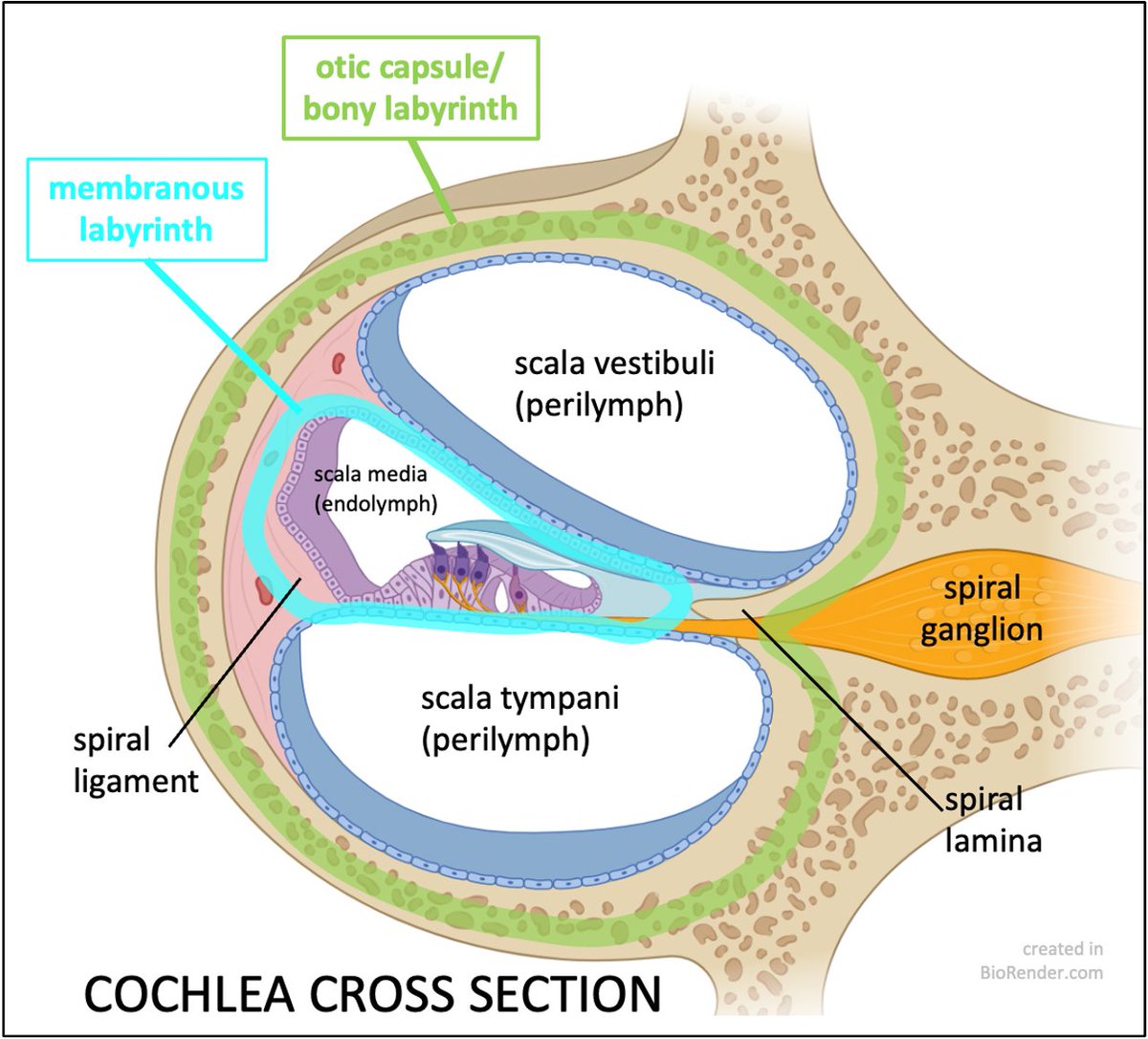
top showcases captivating images of label the parts of the cochlea and spiral organ. galleryz.online
label the parts of the cochlea and spiral organ.
Human ear – Cochlea | Britannica
Auditory Pathways – Reception and Mechanotransduction of Sound – Within …
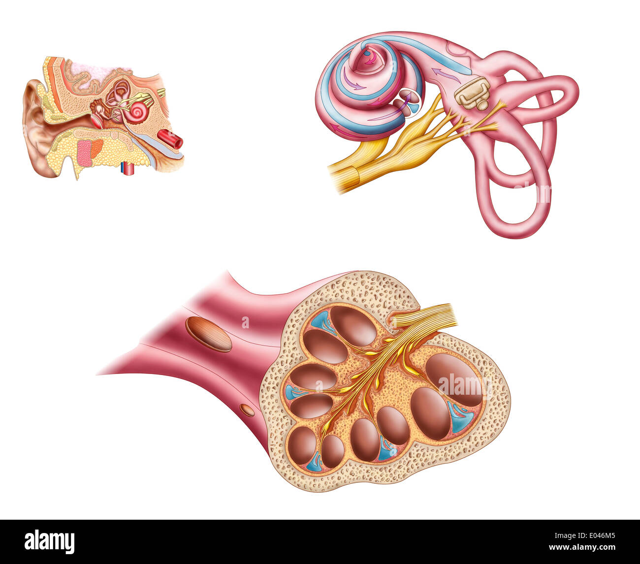
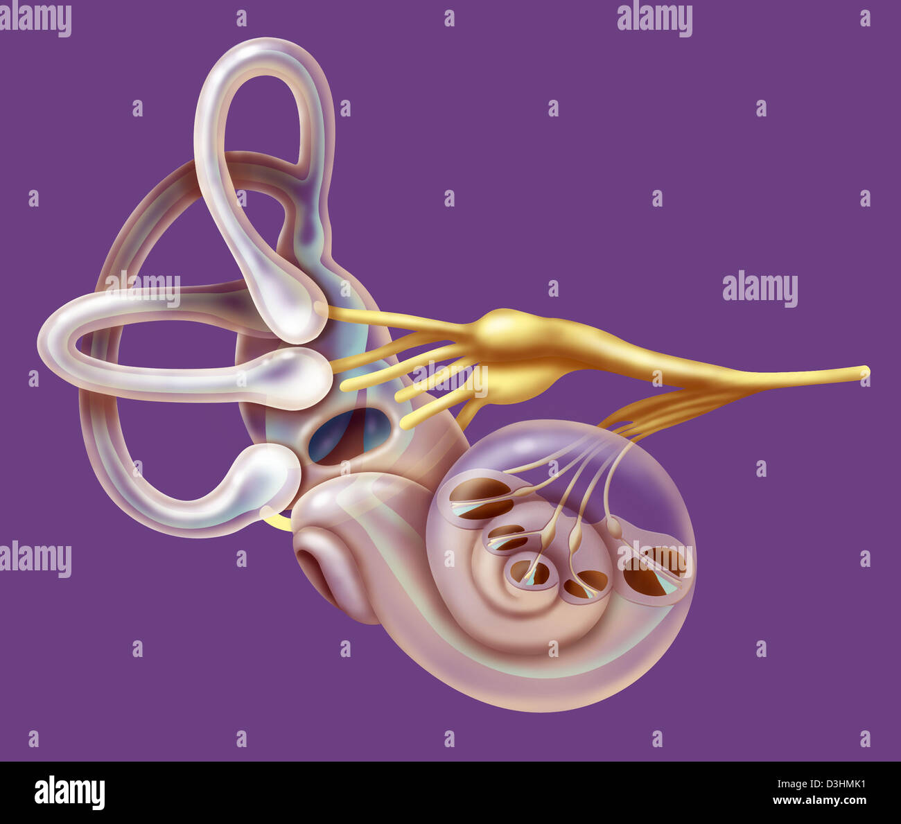
Anatomie der Cochlea des menschlichen Ohres Stockfotografie – Alamy
The Acoustic (Vestibulocochlear) Nerve | Neupsy Key
Vestibular and Auditory Transduction: Hair Cells – Sensory Transduction …
Sensory Processes | Boundless Psychology
Book – Stoehr’s Histology 1-3 – Embryology
Special Senses: Hearing (Audition) and Balance | A & P 1/2
SENSORY TRANSDUCTION – THE NERVOUS SYSTEM – Medical Physiology, 2e …
Photomicrographs of the cochlea and spiral organ of corti (part 1 …
Perspectives for the Treatment of Sensorineural Hearing Loss by …
cochlea and spiral organ of corti Flashcards | Quizlet
Presbycusis – The Lancet
Spiral Organ of Corti 1 Diagram | Quizlet
label the parts of the cochlea Diagram | Quizlet
Frontiers | Resolution of Cochlear Inflammation: Novel Target for …
Cochlear Stockfotos & Cochlear Bilder – Alamy
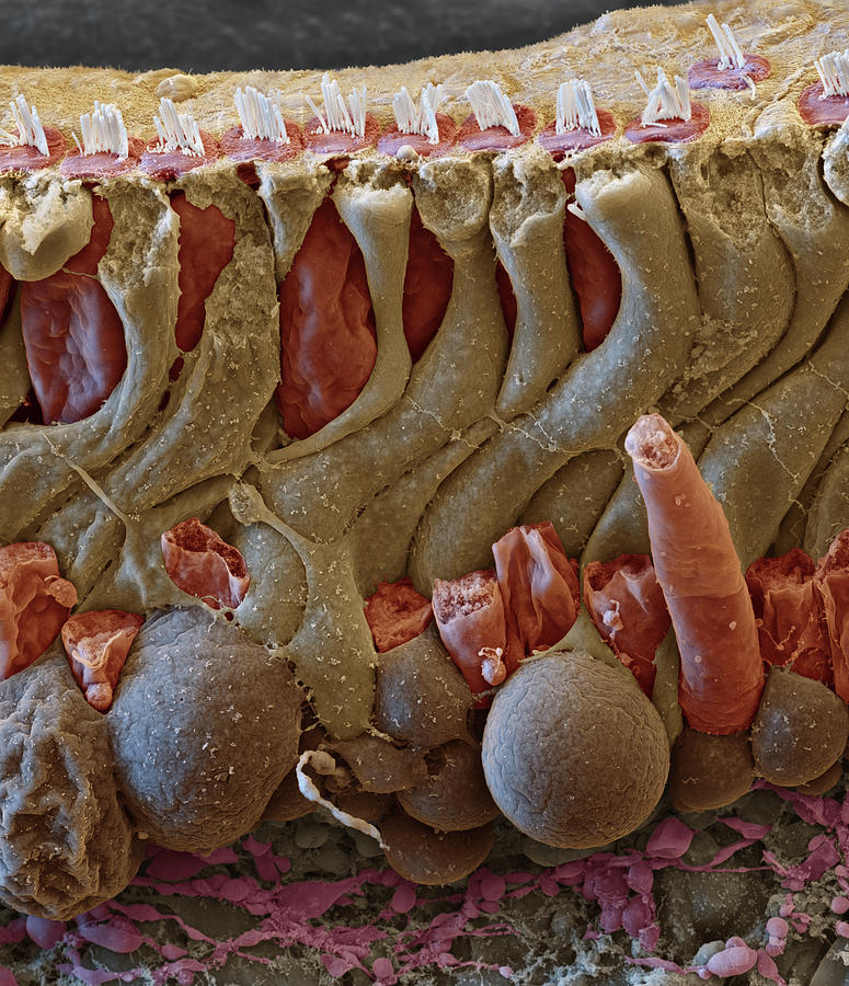
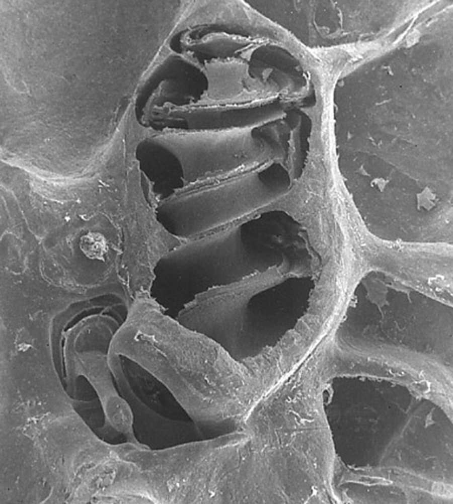
Cochlea, Organ Of Corti Section, Sem Photograph by Oliver Meckes EYE OF …
We Finally Know Why There’s a Bizarre Structure in Our Inner Ears …
Pump up the volume | Issue 22 of Protein Spotlight
An illustration of the cochlea and its tonotopic develo | Open-i
BIO2086L Ch 25 Flashcards | Quizlet
Frontiers | Auditory Nomenclature: Combining Name Recognition With …
Auditory Image Gallery – BrainHQ from Posit Science
100 Pics Body Parts 20 level answer: COCHLEA
Anatomy Of The Ear Stock Illustration – Download Image Now – Anatomy …
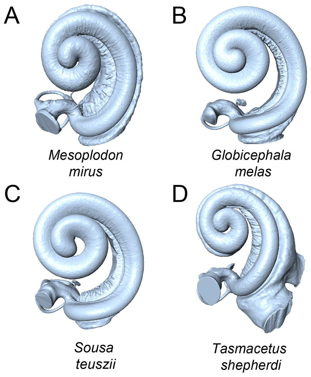
Intraspecific variation in the cochleae of harbour porpoises (Phocoena …
Fluvastatin protects cochleae from damage by high-level noise …
Cochlea Photograph by Mehau Kulyk
Haematoxylin and Eosin (H&E) staining of cochlea se | Open-i
Intraspecific variation in the cochleae of harbour porpoises (Phocoena …
Cochlear Implant Speech Processor – MATLAB & Simulink
Section Through the Turn of the Cochlea
Cochlear Receptors
Pathology Outlines – Anatomy
Cross Section of Cochlea
Structure of the Cochlea and Spiral Organ. The cochlea exhibits a snail …
Answers to this Module
Cochlea and Cochlear Receptors | Hearing health, Biology classroom …
Cochlea | Membrane, Endocrine, Supplemental instruction
Diagram of a cross section of the coiled cochlea | Download Scientific …
DS 10 Section through the Central Spiral of the Cochlea – Biomedical Models
1 Schematic drawing of a cross-section of the cochlea showing the organ …
l113_5_cochleaanatomy
The Ear and Eye | Veterian Key
Cross section of the cochlea. The organ of Corti is located on the …
l113_6_organofcortianatomy
Biology2404 A&P Basics
Potassium Ion Movement in the Inner Ear: Insights from Genetic Disease …
Biology2404 A&P Basics
3 A. Membranous Labyrinth: Showing Scala vestibuli with perilymph …
Inner ear – Auditory Science lab
Inner ear – Auditory Science lab
What is Triformance, and How Does it Work? | The MED-EL Blog
cochlea (conceptual documentation)
5 Cross-section of the cochlea | Download Scientific Diagram
Anatomy of the organ of Corti, part of the cochlea of the inner ear …
[Solved] Label the anatomy of the cochlea…. | Course Hero
Schematic diagram of a cross-section through one turn of the mammalian …
Cochlea Diagram Anatomy | Health | Pinterest
Spiral Ligament of Cochlea | Semantic Scholar
Diagram of a cross section of the coiled cochlea | Download Scientific …
Schematic demonstrating the basic anatomy of the cochlea. A. Cross …
2: Figure showing an unwound cochlea. Due to the physical properties of …
3: A cross section of the cochlea. Inner hair cells detect the movement …
IAS (Admin.) Mains Zoology: Questions 1 of 48 – DoorstepTutor
Human cochlea anatomy stock illustration. Illustration of ossea – 66046862
Cell types present in the cochlea and the endogenous or transgenic …
2: Schematic representation of the uncoiled cochlea. Reproduced from …
Chapter 16 Earth Science Quizlet – The Earth Images Revimage.Org
Compartments of cochlea with organ of Corti and stereochilia basic …
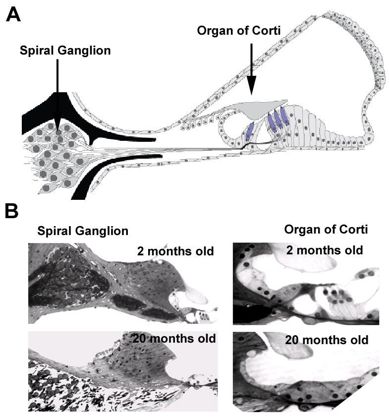
Why do hair cells and spiral ganglion neurons in the cochlea die during …
Electron microscopy – Auditory Science lab
Spiral Organ Corti High Resolution Stock Photography and Images – Alamy
Cross-section of the cochlea with enlarged organ of Corti [40 …
Structure of Organ of Corti | Download Scientific Diagram
Anatomy of the Cochlea. Cartoon illustration of the cochlea. Panel a. A …
Schematic drawing of the cochlea in cross section. The cochlear duct …
Brain Function and Auditory Image Gallery – DynamicBrain
64 best images about Illustrative & Graphic Art on Pinterest | Pelagic …
Schematic representation of the cochlea and the afferent innervation of …
1. Schematic of the auditory system with its primary components …
Inner ear. Cochlea stock image. Image of tissue, spiral – 117240643
Tectorial Membrane Photos and Premium High Res Pictures – Getty Images
Anatomical details of inner ear, cochlea and organ of Corti, the sense …
Cross section of the cochlea (A) and the structure of the organ of …
Meniere’s disease – Audiology BSc | enteducationswansea
Schematic diagram of a cross section of the cochlear duct showing the …
Light microscopy – Auditory Science lab
Human Ear Anatomy – Parts of Ear Structure, Diagram and Ear Problems
Cóclea da disseção ilustração stock. Ilustração de exterior – 32128291
Special Senses: Hearing. – The Biotech Notes
Thread by @aaronrutman on Thread Reader App – Thread Reader App
Otolaryngology: Diseases Of The Head And Neck | Basicmedical Key
Ljudv gors transduktion
We extend our gratitude for your readership of the article about
label the parts of the cochlea and spiral organ. at
galleryz.online . We encourage you to leave your feedback, and there’s a treasure trove of related articles waiting for you below. We hope they will be of interest and provide valuable information for you.


