Albums showcases captivating images of echocardiogram pictures of the heart gathered and meticulously curated by the website galleryz.online. Furthermore, you can find more related images in the details below.
Echocardiography showed a small amount of pericardial effusion (PE …
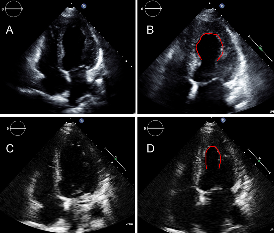
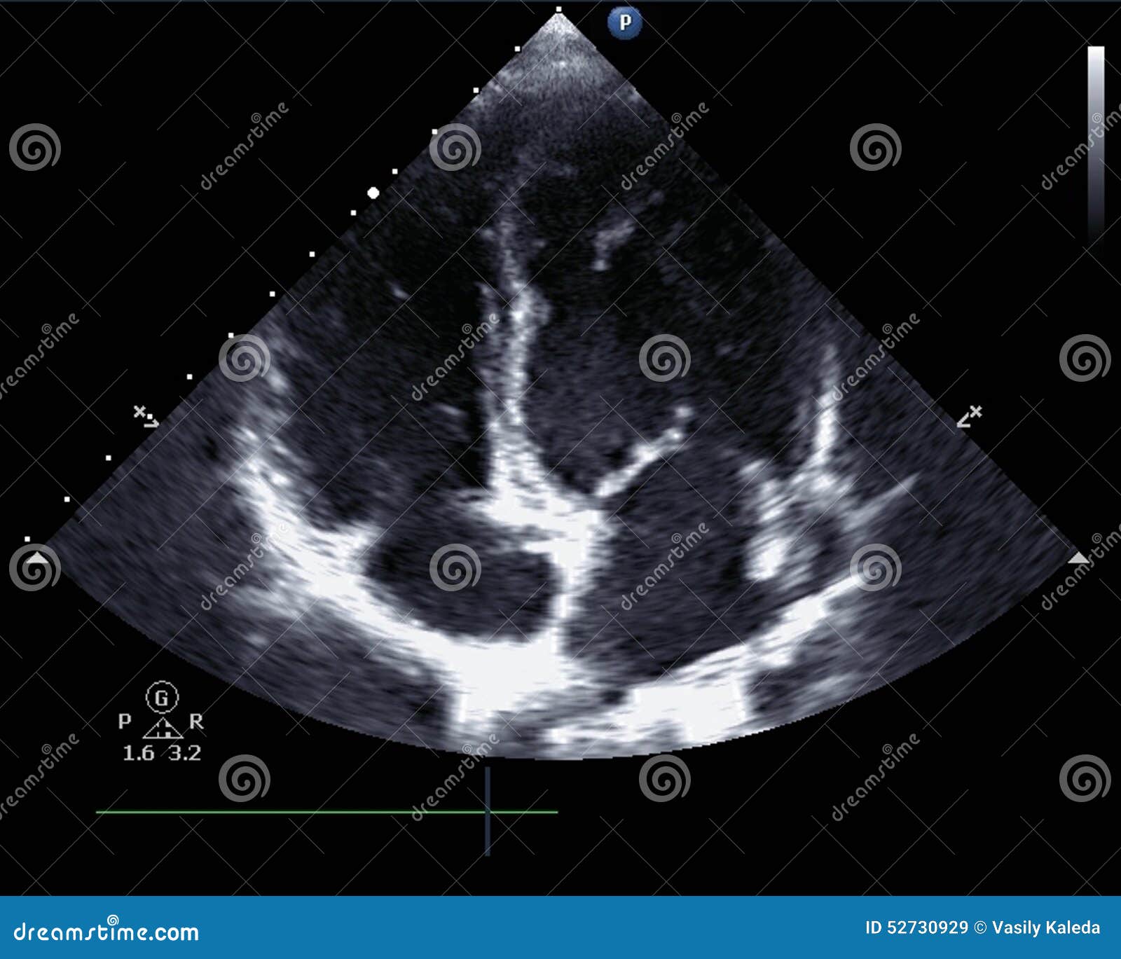
The transthoracic echocardiogram showed akinesia of anterolateral …
Bacterial Endocarditis | Circulation
Making sense of an echocardiogram report – for GPs! — Cardiology Institute
-Transthoracic echocardiogram of a right ventricular aneurysm. Four …
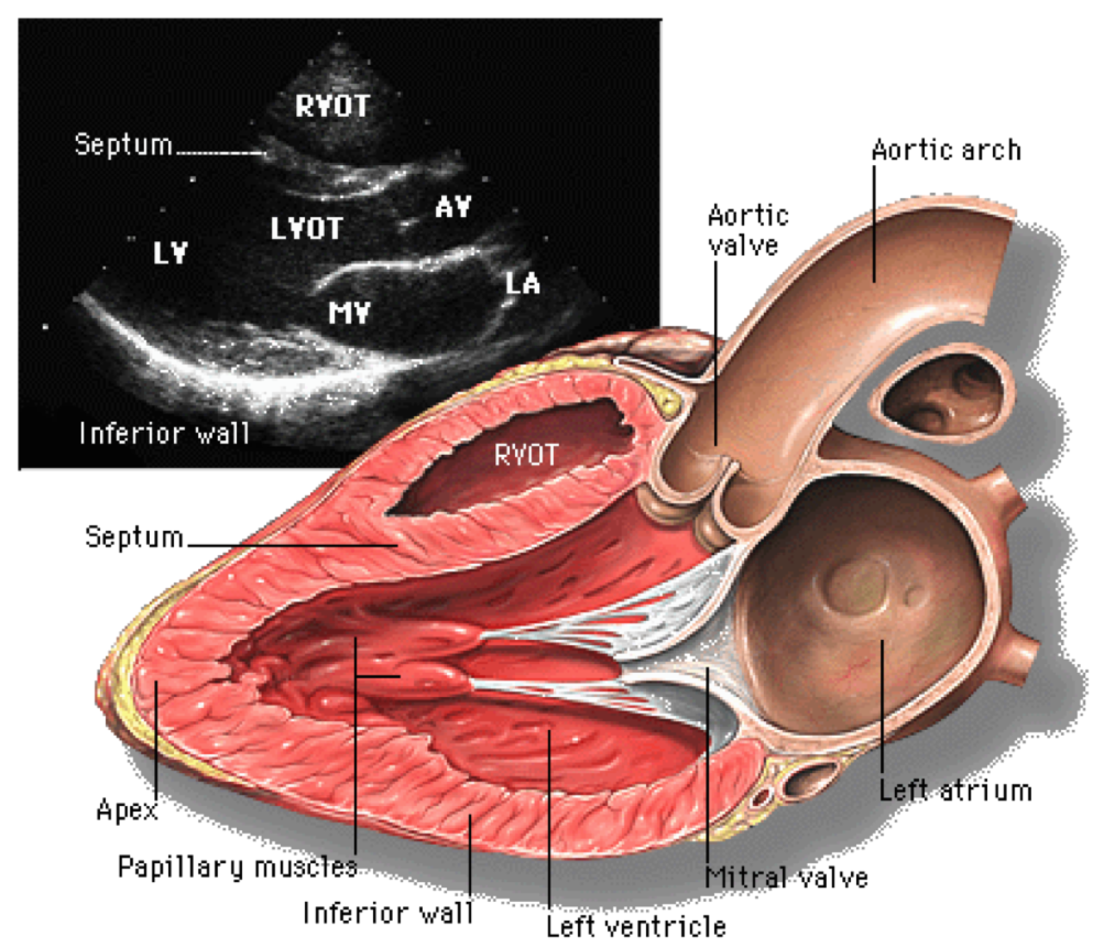
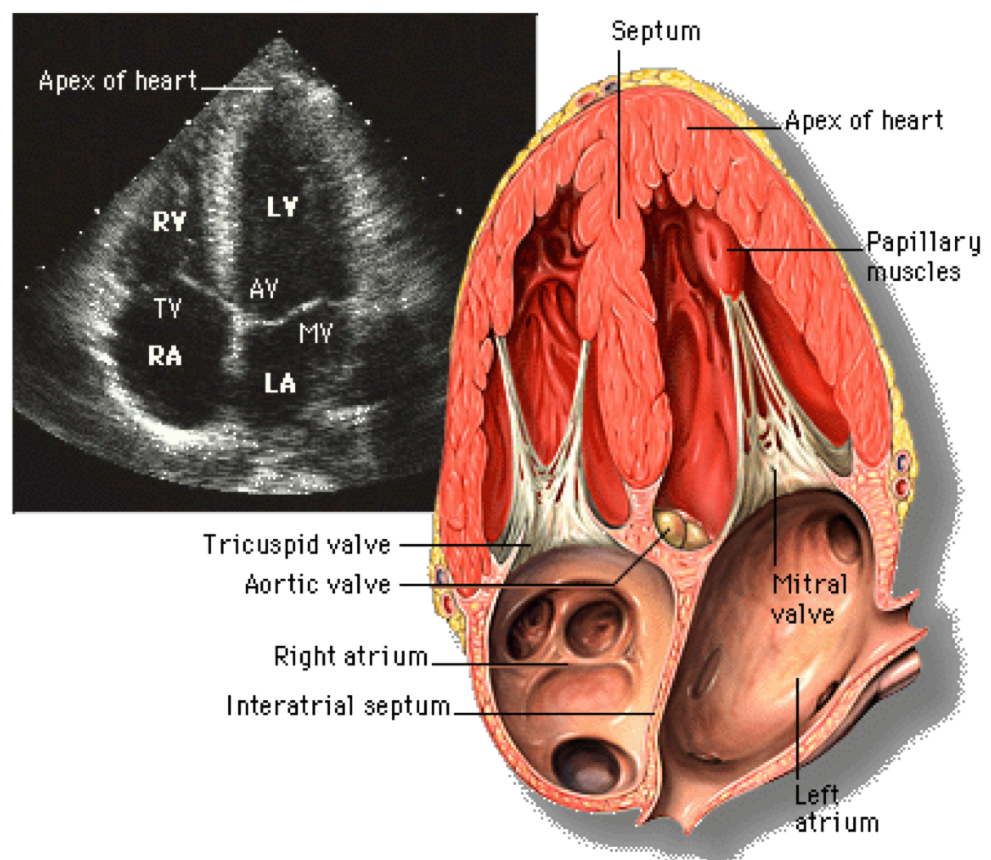
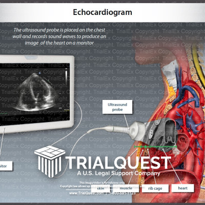
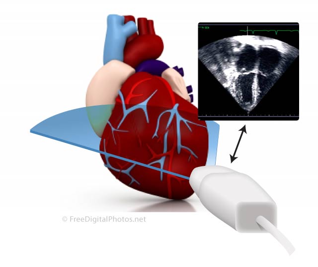
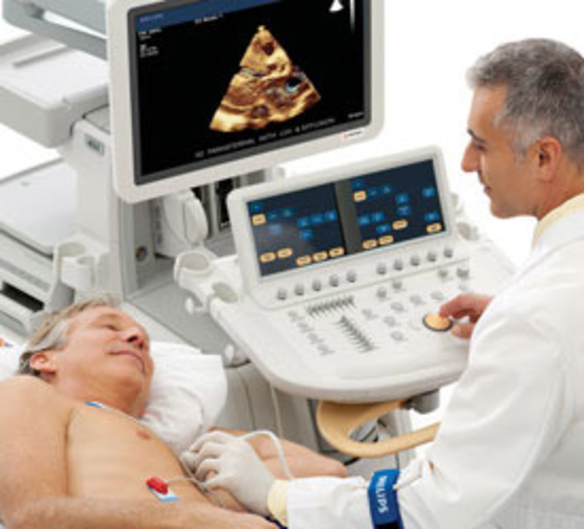
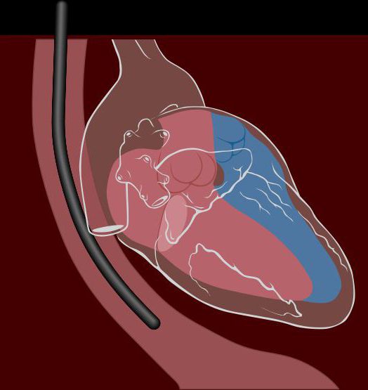
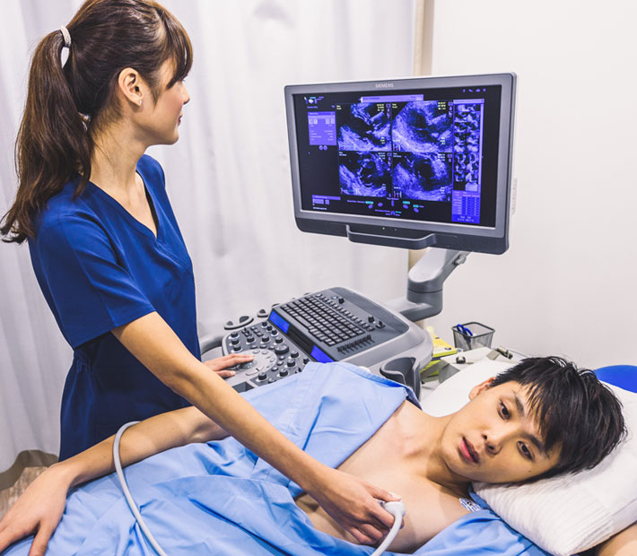
The Echocardiogram – Congenital Heart Disease
Cardiac Stress Testing in the Emergency Department | 2008-09-01 | AHC …
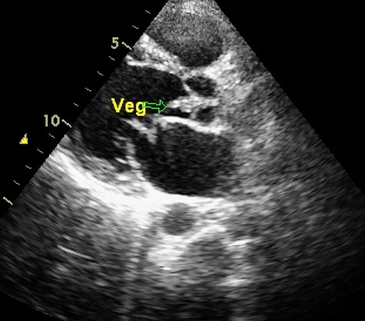
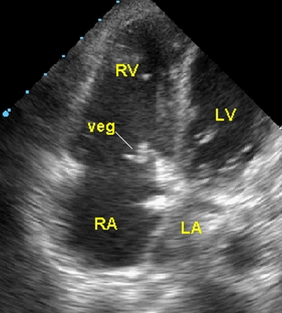
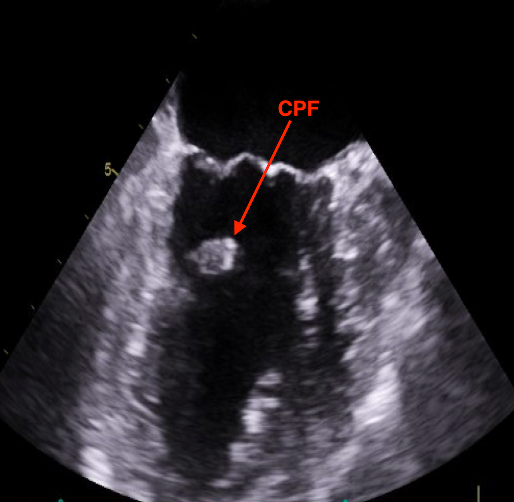
Four chamber view echocardiogram showing large vegetation on the MVL …
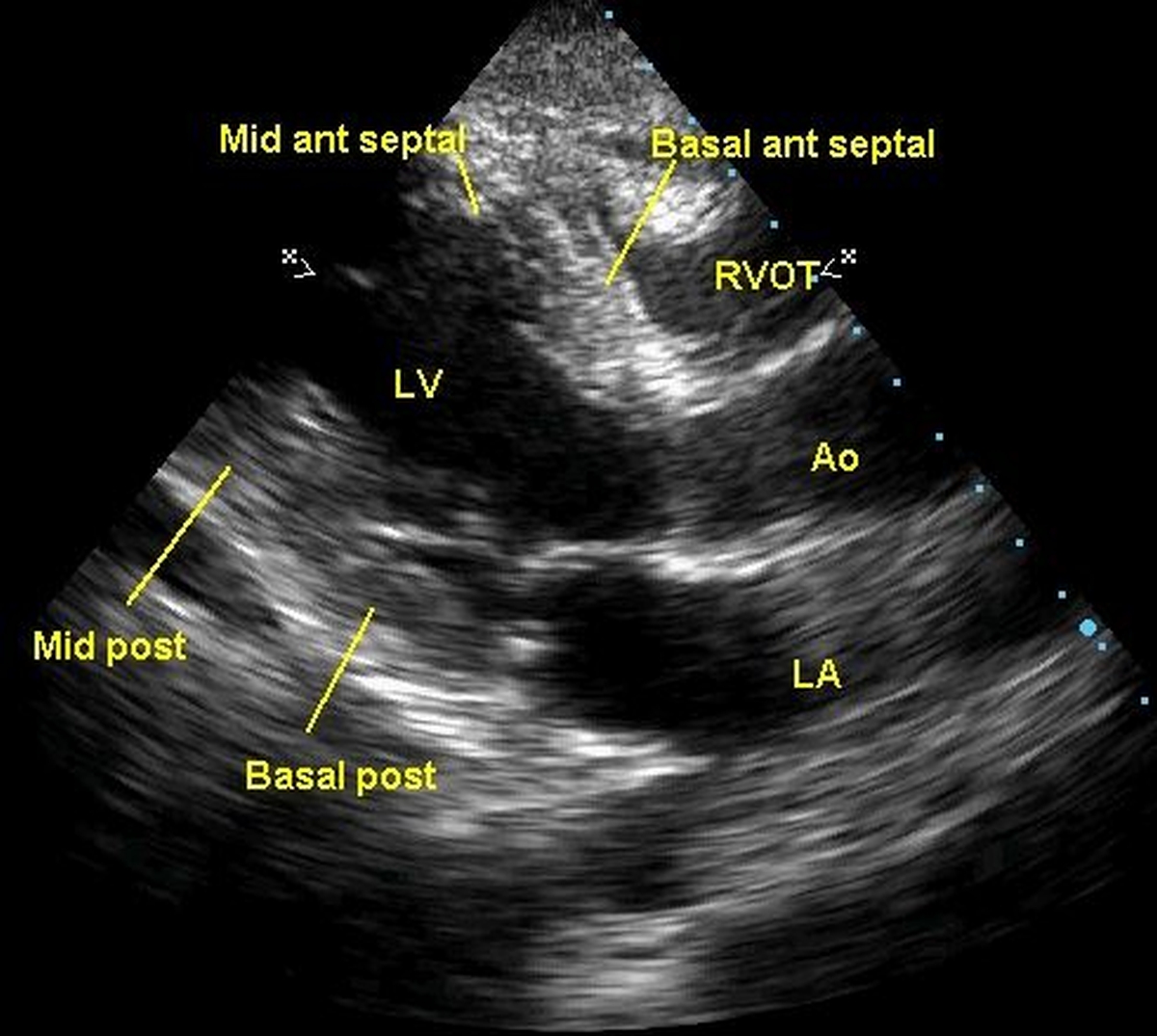
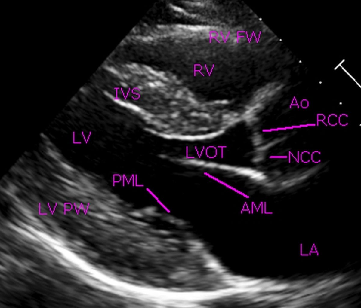
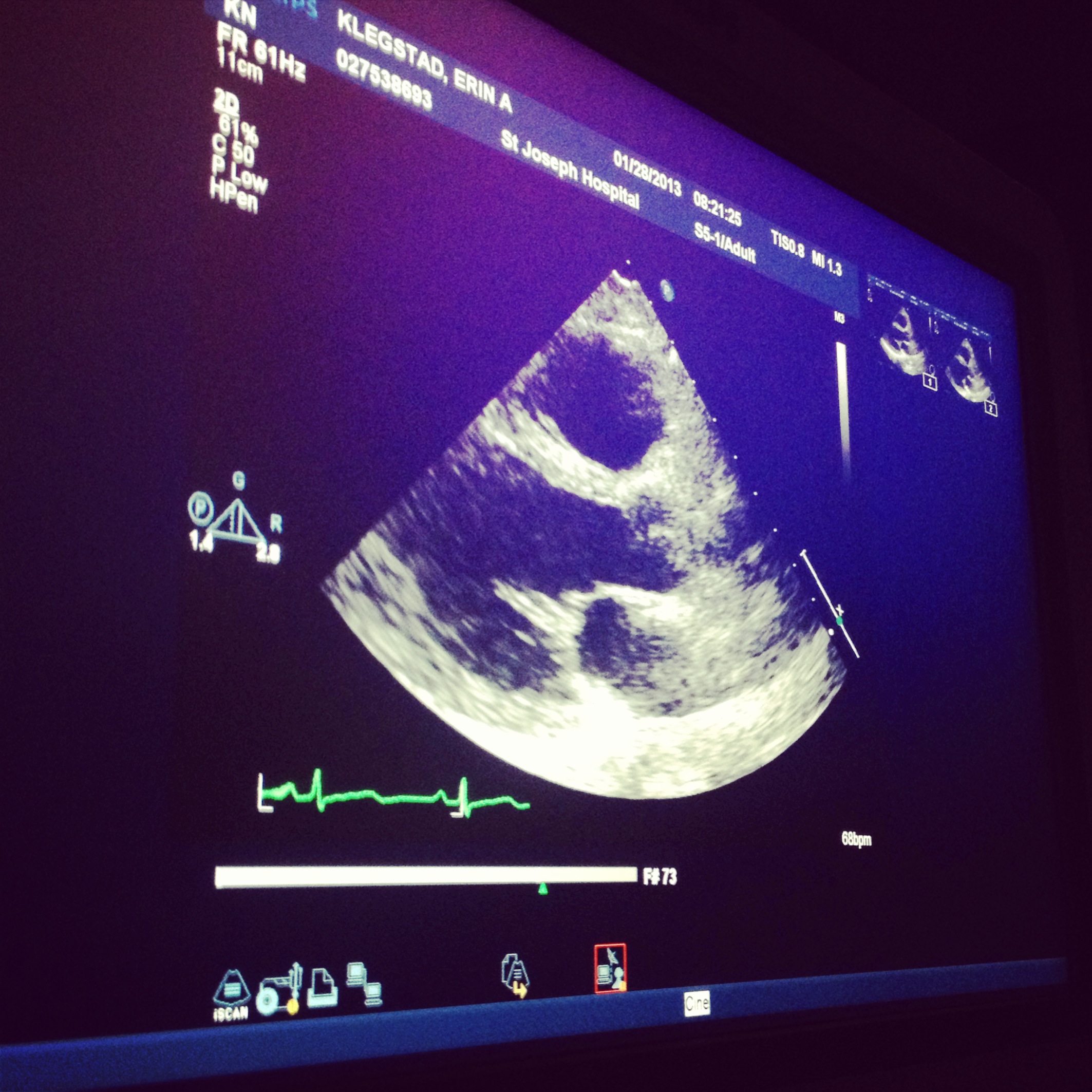
Transthoracic echocardiogram (parasternal long axis view) showing …
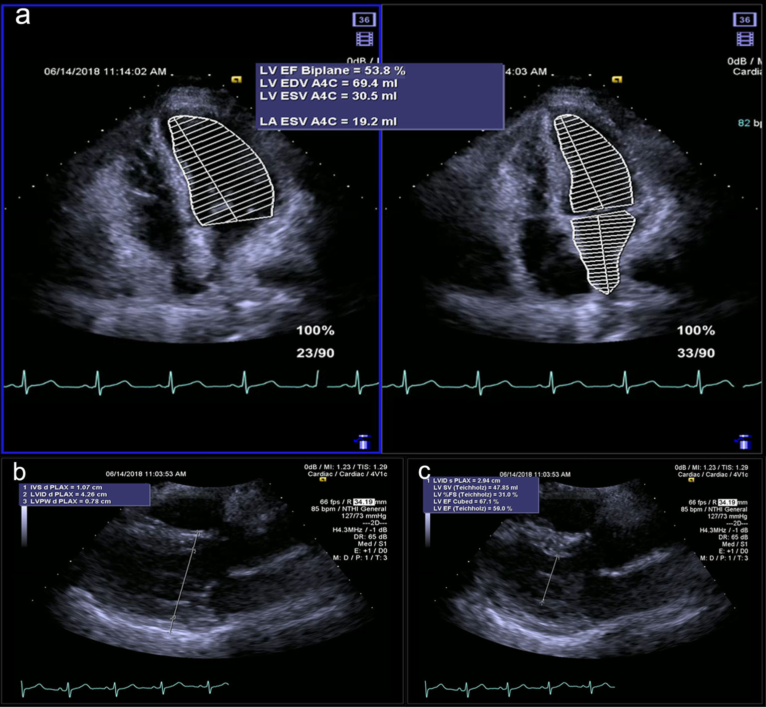
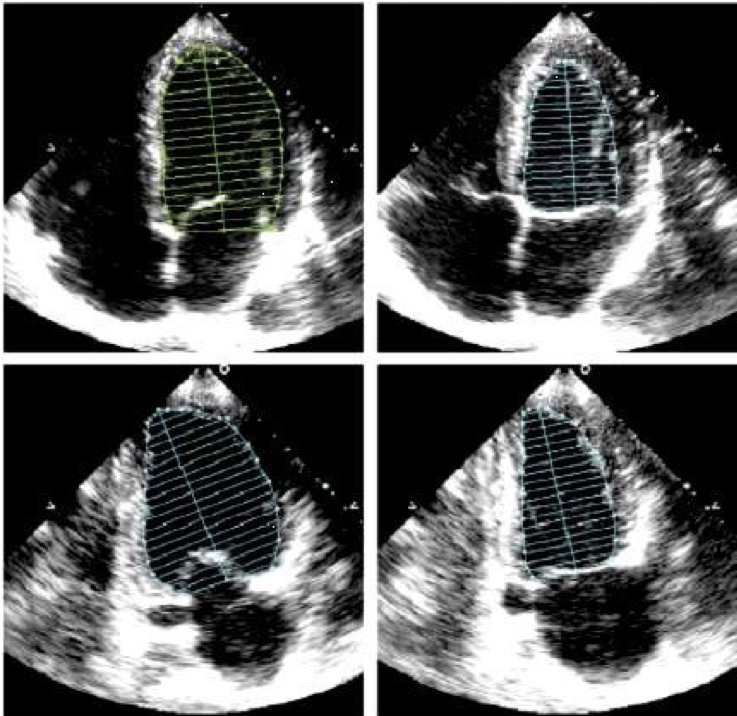
Echocardiographic assessment of left atrial volume index in elderly …
Transesophageal echocardiogram showing possible flail mitral valve and …
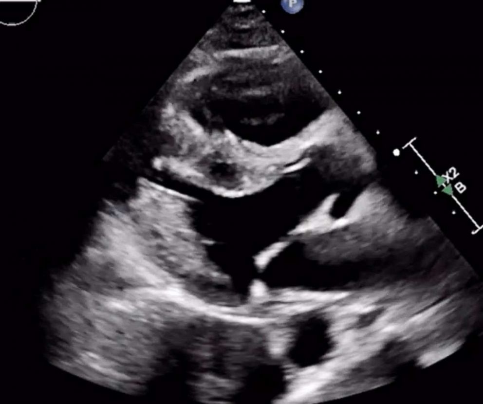
Two-dimensional cardiac echocardiogram. In a subcostal view of the …
Right heart chambers dilatation. (A): Transthoracic echocardiogram …
Echocardiogram demonstrating the presence of vegetation in the …
(PDF) Constrictive pericarditis
Echocardiograms (a) from 2 patients with heart failure, mild (left) and …
Echocardiogram, Heart Ultrasound Color Line Icon. Isolated on White …
Pin on Work stuff
Transesophageal echocardiogram of a juxtaarterial ventricular septal …
CTEPH.com | The Importance of Echocardiography
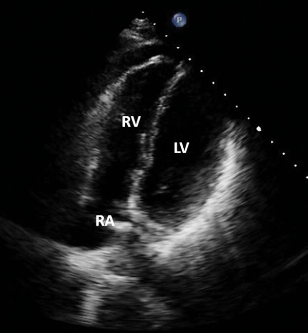
Transthoracic echocardiogram of the left and right ventricles. The left …
Echocardiogram showing coarctation of aorta distal to left subclavian …
Transthoracic echocardiography. White arrow indicates apical …
We extend our gratitude for your readership of the article about echocardiogram pictures of the heart at galleryz.online. We encourage you to leave your feedback, and there’s a treasure trove of related articles waiting for you below. We hope they will be of interest and provide valuable information for you.