Collection showcases captivating images of how do the highlighted valve cusps function during a ventricular contraction? gathered and meticulously curated by the website galleryz.online. Furthermore, you can find more related images in the details below.
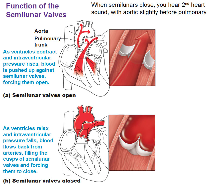
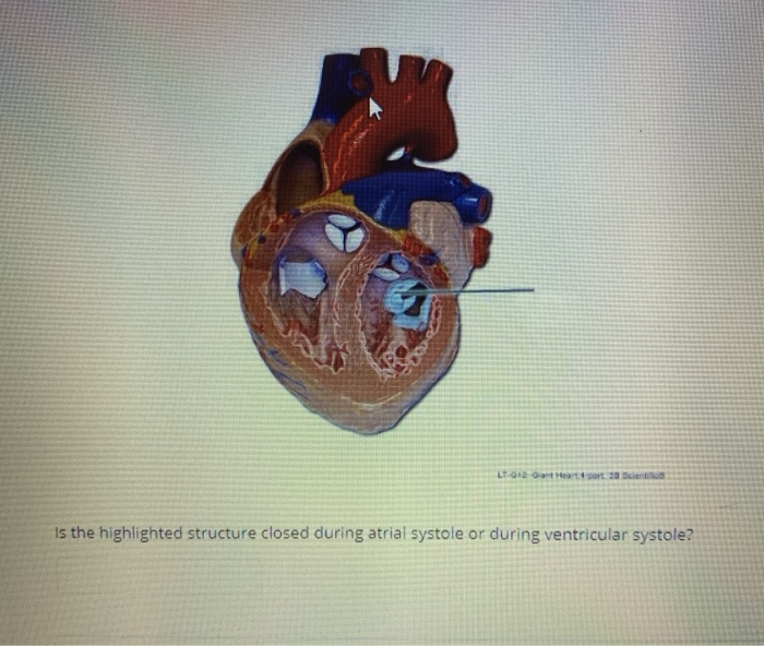
how do the highlighted valve cusps function during a ventricular contraction?
 Which Heart Valve Has Two Cusps – Add Tech Curry
 Left Atrioventricular Valve | Anatomy, Heart valves, Cardiac
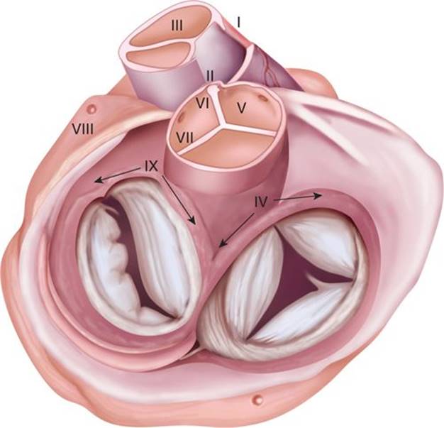
 | Spatial relationship between the three cusps of the aortic valve and …
 Pin on Medschool Resources
 VENTRICULAR TACHYCARDIA AND CARDIAC ANATOMY: AORTIC CUSP – Color Atlas …
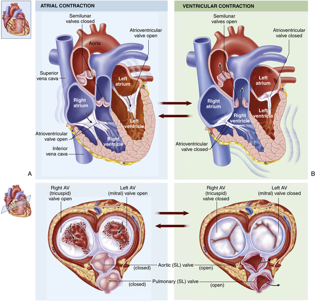
 OpenStax: Anatomy and Physiology | CH19: THE CARDIOVASCULAR SYSTEM: THE …
 Electrophysiological Characteristics of Focal Atrial Tachycardia …
 The atrioventricular valve’s are found between the atria and the …
 Structure and Function of the Cardiovascular and Lymphatic Systems …
 cusps of aortic valve face septum an anatomical plate a human heart …
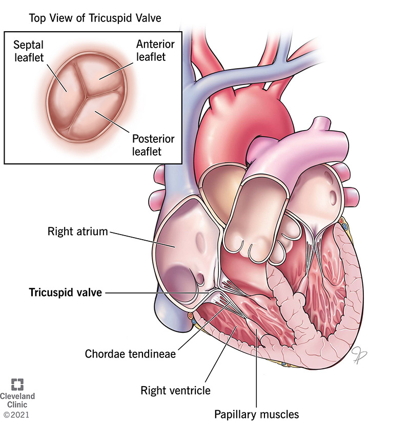 Tricuspid Valve: Overview, Function and Anatomy
 The valve present between the left auricle and left class 11 biology CBSE
 Atrioventricular v’s | definition of atrioventricular v’s by Medical …
 Anatomic positions of ventricular septal defects. The major anatomic …
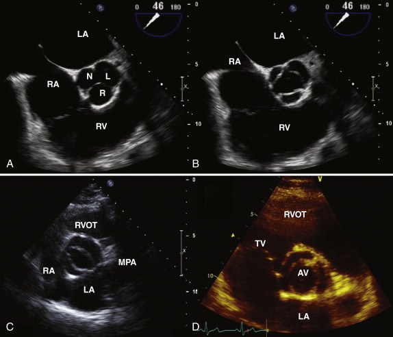
 Short axis view showing the aortic valve as a trileaflet structure with …
 Association Between Bicuspid Aortic Valve Phenotype and Patterns of …
 Mitral Regurgitation Article
 Top, normal tricuspid valve with anterior, posterior, and septal …
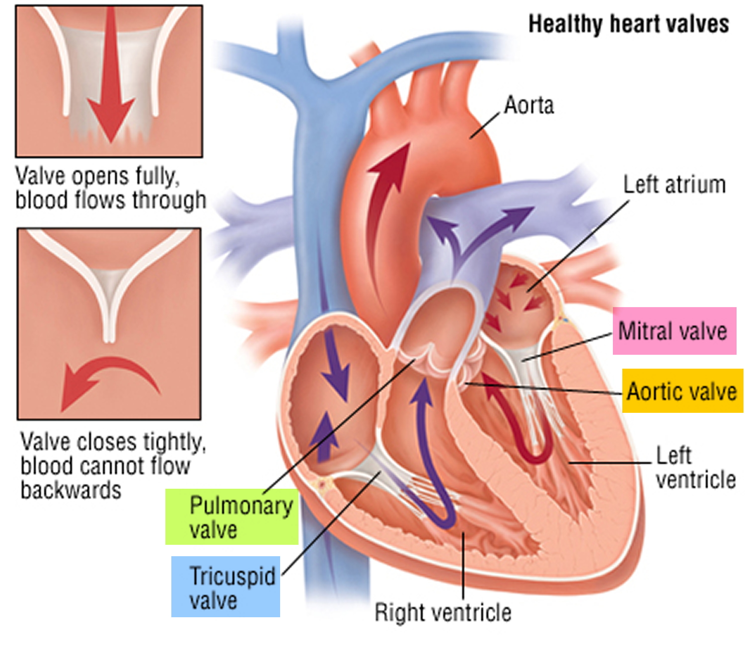
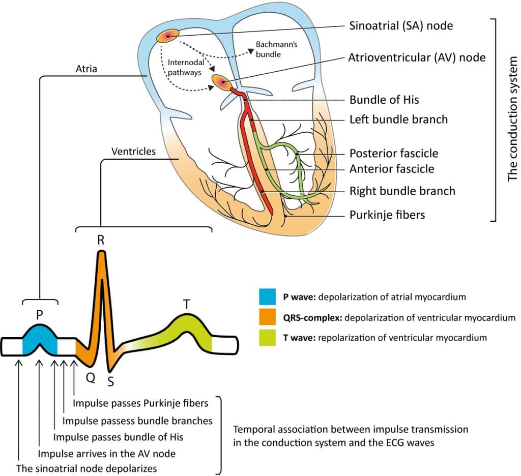
 Normal Heart Function and Normal Heart Rhythm
 TTE parasternal short axis views of three aortic valves with leaflets …
 Echocardiogram, Transesophageal, Transthoracic, Stress Echocardiogram
 The Heart as a Pump | Basicmedical Key
 Shape of anterior, posterior, septal cusp of tricuspid valve | Download …
 Atrial ventricular accessory pathways may occur anywhere along the …
 View Image
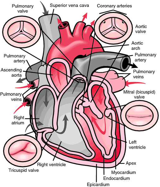
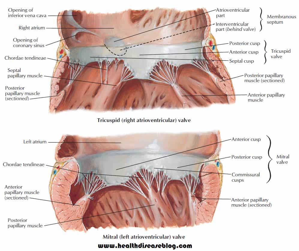
 Heart valves. | Human heart anatomy, Human anatomy and physiology …
 Aortic valve short-axis view (ac, acoronary; lc, left coronary; rc …
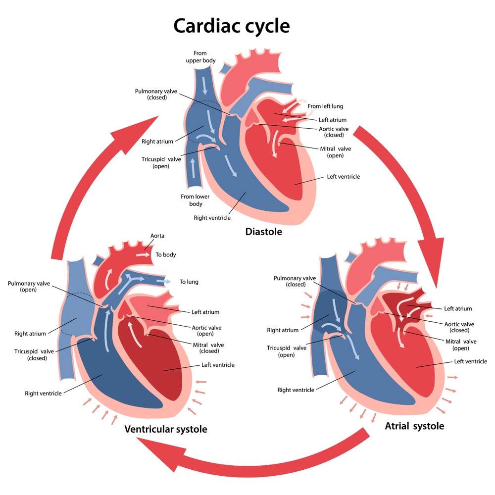
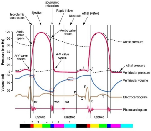 The entire two cardiac cycle diagram, which contains in | Open-i
 Av Valves of Heart submited images | Pic2Fly
 (A) Long-axis TOE view showing the left ventricular outflow tract …
 TEE image of the aortic valve in short-axis view demonstrating four …
 Phases of the Cardiac Cycle Diagram | Quizlet
 Feature of PVC on the surface 12-lead ECG that originated from the …
 Echocardiography analysis of the aortic valve structure and function in …
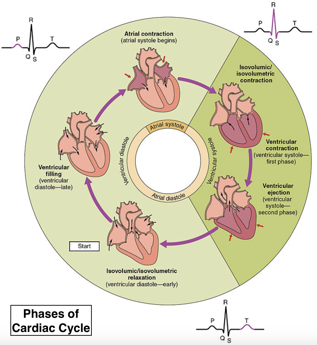 Define the cardiac cycle Describe various events of class 11 biology CBSE
 Twelve-lead electrocardiogram morphology of different sites of origin …
 Aortic valve function. A and B: aortic cusp separation (ACS). C …
 What Is Heart Valve Disease?
 Diagram illustrating the components of the aortic valve complex. Note …
 deflection point generated by ventricular repolarization | wave …
 Pin by Melissa Blaker on Cardiac Surgery | Medical knowledge …
 19.3 Cardiac Cycle – Anatomy & Physiology
 Anatomy of the Aortic Valvar Complex and Its Implications for …
 Chapter 28 – Cardiac Cycle | Anesthesia Key
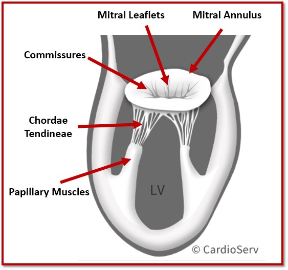 Papillary Muscle Names / Canine papillary (heart) muscle | 2000 …
 Cardiac cycle – Isovolumic Contraction In cardiac physiology …
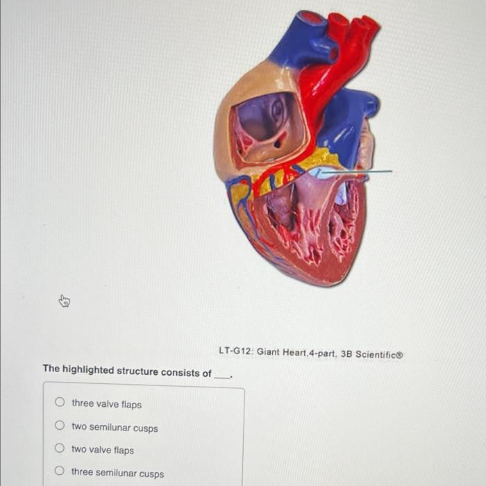 Solved Model courtesy of Denoyer-Geppert, www.denoyer.com | Chegg.com
 Dimensions of the aortic valve cusps, whereby R is the radius of the …
 Aorto-right atrial and right ventricular fistulae: a very rare …
AQUARIUS /PISCES CUSP~ALL THE HARD WORK IS PUSHING YOU TOWARDS BIG REWARDS!!🏆
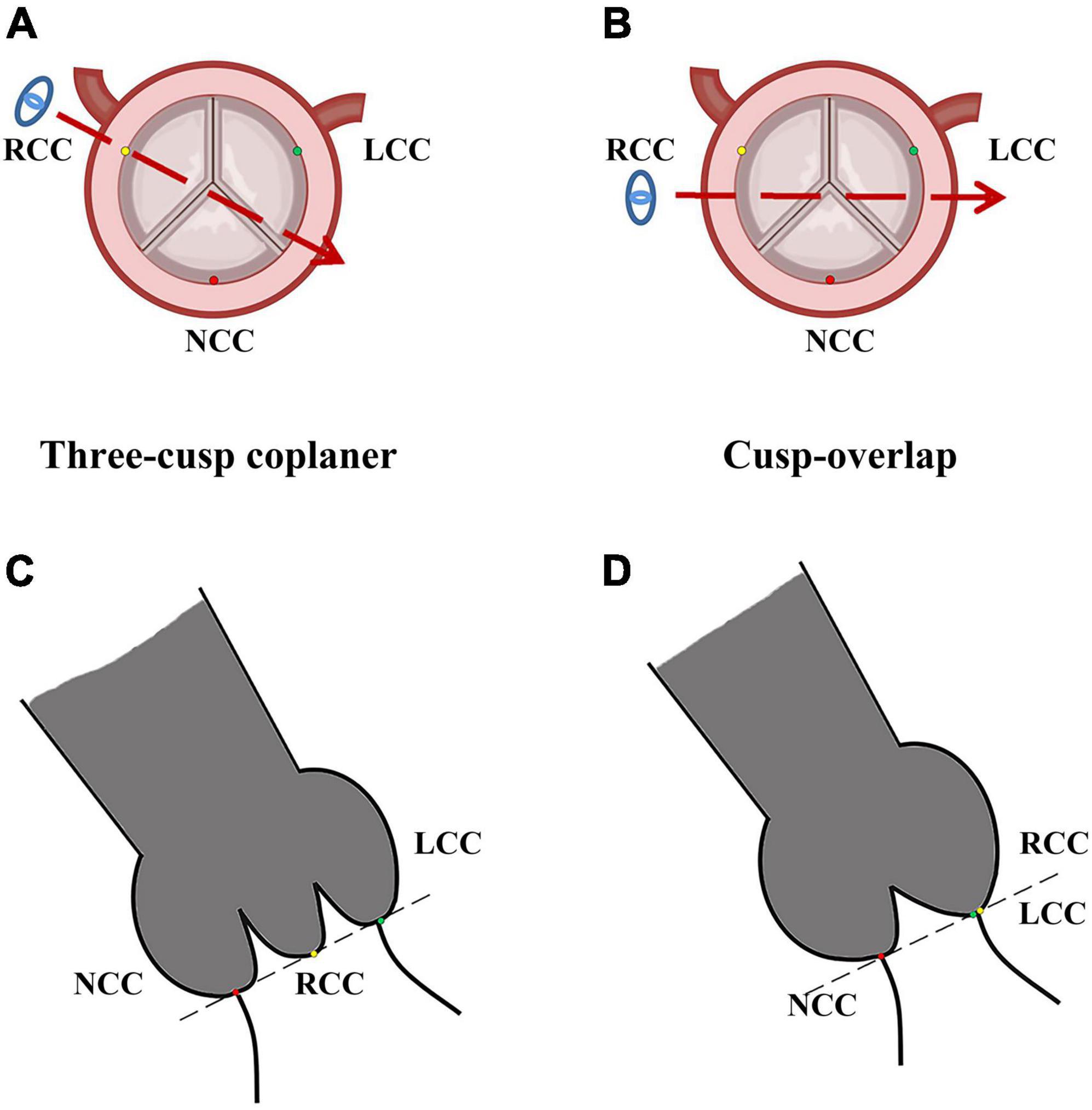 Frontiers | Comparison of cusp-overlap projection and standard three …
 Illustration of a prolapsed mitreal valve in the heart | Mitral valve …
 The safely resectable region of the interventricular septum, mostly …
 Cardiac Function | Basicmedical Key
 😱 When contraction occurs. What happens during uterine contraction …
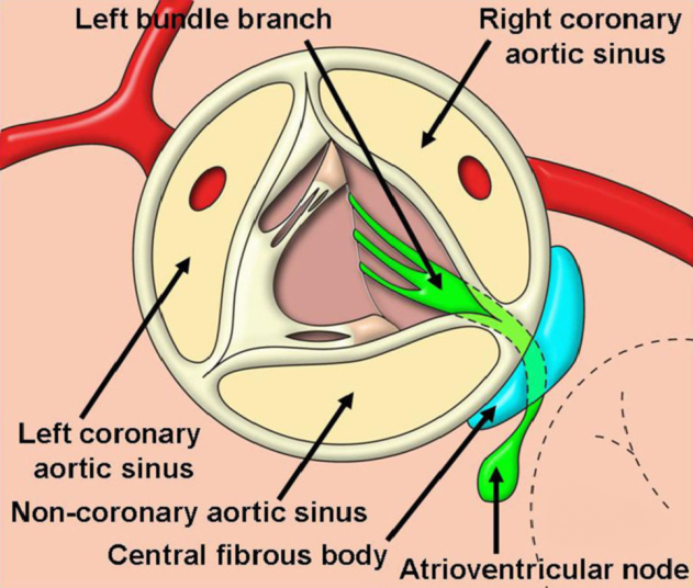
 Schematic showing the central location of the aortic valve and the …
 Deployment of the Evolut PRO + transcatheter aortic valve system using …
 a Successful ablation of a PVC originating from the right coronary cusp …
 Native aortic valve cusps after surgery excision. The laceration of the …
 Difference Between Mitral Valve and Tricuspid Valve | Compare the …
 Right Ventricular Anatomy
 Ventricular Pressure-Volume Relationship: Preload, Afterload, Stroke …
 Comprehensive 4-stage categorization of bicuspid aortic valve leaflet …
 Schematic representation of a bicuspid aortic valve. (a) Transverse …
 Anatomy Choice of surgery Aortic Valve Repair Aortic Valve Replacement …
 Anatomy and Function of Normal Aortic Valvular Complex | IntechOpen
 During Ventricular Systole What Closes the Av Valves
 Left ventricular diastolic function – ECG & ECHO
:watermark(/images/logo_url.png,-10,-10,0):format(jpeg)/images/anatomy_term/valvula-semilunaris-sinistra-valvae-aortae/DKmvDEJznLxbTwH8NFJ3dA_Valvula_semilunaris_sinistra__Valva_aortae__1.png) Left semilunar cusp of aortic valve (Valvula semilunaris sinistra …
Left semilunar cusp of aortic valve (Valvula semilunaris sinistra …
 VENTRICULAR TACHYCARDIA AND CARDIAC ANATOMY: AORTIC CUSP – Color Atlas …
 Cardiac Cycle – Definition, Phases and Quiz | Biology Dictionary
 Unusual Outflow Tract Ventricular Tachycardia – Cardiac …
 Valves: Echocardiography | Radiology Key
 Normal aortic valve-short axis. Two-dimensional (2-D) and… | Download …
 Gross Anatomy Glossary: Heart Valves | Draw It to Know It
 Systolic Anterior Motion (SAM) Of The Mitral Valve – Left Ventricular …
 Aortic Valve | Anesthesia Key
 Solved: Identify The Highlighted Vessel. Identify The High… | Chegg.com
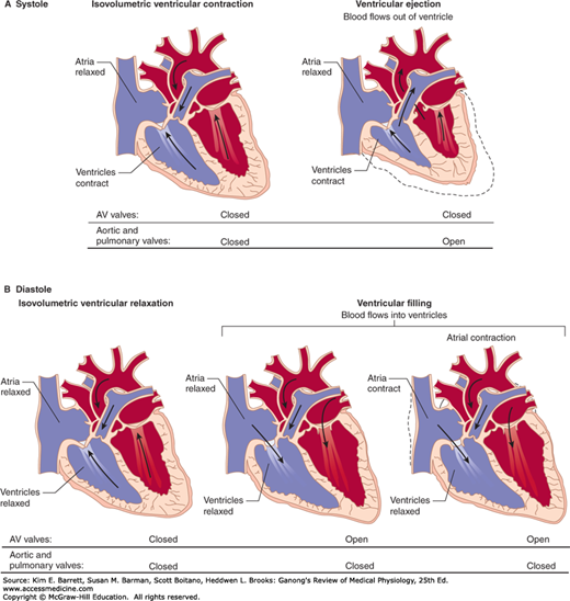
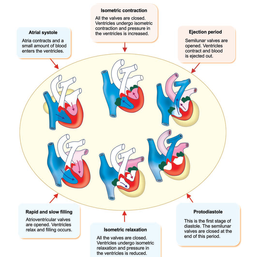 Cardiac Cycle VENTRICULAR EVENTS
 Ssurvivor: Malar Mitral Facies
 Clinical electrocardiography and ECG interpretation – ECG interpretation
 The image shows the undersurface of the closed aortic valve of a …
 Pulmonary valve cusp augmentation with autologous pericardium may …
 The parasternal short-axis view on the aortic valve at two-dimensional …
 Pace mapping and ablation of premature ventricular contraction (PVC …
 Shape of anterior, posterior, septal cusp of tricuspid valve | Download …
 Coronary anatomy as related to bicuspid aortic valve morphology | Heart
 Pin on Cardiology
 heart valve sound locations – Google Search | Nursing school notes …
 Pulmonary Valve | Pulmonary, Heart care, Anatomy
 Aortic Valve | Anesthesia Key
 The Ross Procedure – Operative Techniques in Thoracic and …
 TTE short-axis view at the level of the prosthetic aortic valve. (a …