Collection showcases captivating images of how to make copies of ultrasound pictures galleryz.online
how to make copies of ultrasound pictures
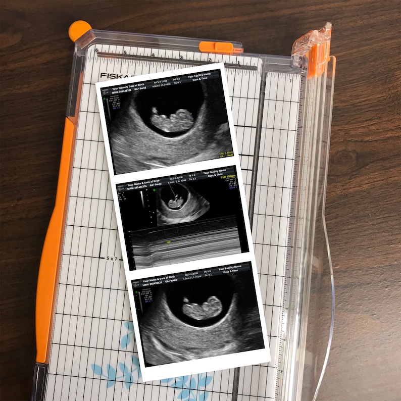
Fake Baby Ultrasound Sonogram Prank | WEIRD THINGS TO BUY
Sonography. Ultrasound. | Medical ultrasound, Ultrasound technician …
3 Reasons We Offer Free Ultrasounds | Selah
VIDEO
How to become a pediatric sonographer and/or a neurosonographer *ultrasound career*
APA ITU ULTRASOUND SCAN? – Ultrasound Scan Baby Malaysia – DR SITI SCAN
Fake Ultrasounds Fake Sonogram Fake Your Baby Daddy Belly Bump
Custom Prank 2D 1 PHOTO Pregnancy Ultrasound Sonogram REAL ULTRASDOUND …
Ultrasound scans at 8 weeks pics.
Ultrasound images of a normal fetus (a) and a fetus with trisomy 21 (b …
Ultrasound Pictures: Ultrasound Pictures By Month
Ultrasound showed ventriculomegaly, agenesis of corpus callosum and …
O Baby! Murfreesboro, TN Gender Determination. 3D/4D Ultrasounds …
Abdominal Ultrasound showed severely dilated stomach. | Download …
Liver Anatomy and Protocol basics – Sonographic Tendencies | Liver …
3D Ultrasound | 4D Ultrasound Images & Photos – UC BABY
www.ababyvisit.com 3d ultrasound of baby | 3d ultrasound, Ultrasound …
How To Tell Gender On Ultrasound At 12 Weeks
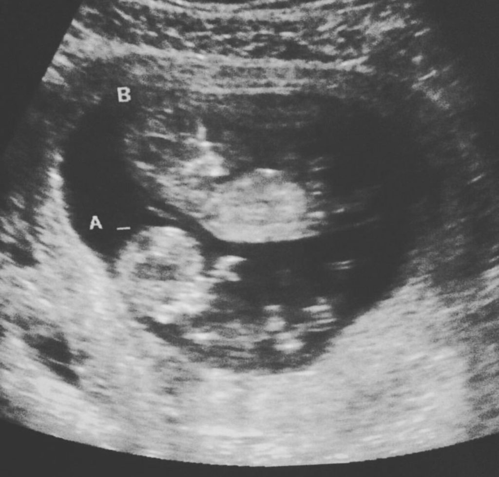
10 week ultrasound – Winnie
Patient 2: Pelvic Ultrasound: didelphys uterus with two endocervical …
(a) Transvaginal ultrasound scan (longitudinal view) showing free fluid …
SONOGRAPHY PRINCIPLES – MK Ultrasound Training center
Ultrasound image of a left axillary vasculature. | Download Scientific …
Fake Ultrasound Maker – Fake Pregnancy Ultrasound Maker with Instant …
Pin by 3D 4D Baby Breath Ultrasound S on Customer 3D Pictures in Black …
Transverse transvaginal ultrasound image showing (U) uterus, (C …
Acute Appendicitis | Medical ultrasound, Ultrasound school, Ultrasound …
9 Month Pregnancy Baby Ultrasound – babypregnancy
Pin by Jennafer Evans on Ultrasound | Ultrasound sonography, Medical …
The Radiologist on Instagram: “LIVER ULTRASOUND 👨🏽💻Ultrasound of …
Early dating scan 4 weeks | Early Ultrasound Scans weeks 7 8 9 10 week …
7.5 Grey-scale ultrasound features of normal thyroid gland and its …
Fake Ultrasound Pictures Prank Ultrasound Picture Digital | Etsy
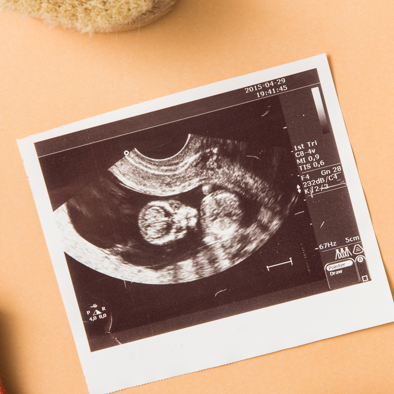
Pictures of an ultrasound framed simply like this make such a sweet …
Dog Ultrasound – The O Guide
(A) An ultrasound image of the urinary bladder (transverse scan …
Abdominal ultrasound images of the pancreatic region in representative …
The Doppler ultrasound showed partial thrombosis of the superior …
Patterns of nerve ultrasound changes in peripheral neuropathy (PN …
Gray-scale ultrasound image of the pancreas shows an isoechoic lesion …
Amazon.com: Sonogram Baby Ultrasound Picture Frame Shower Gift …
Scrotal ultrasound: sagittal image of the normal testicle, showing …
Pin on Scrapbooking
Prenatal ultrasound: (A) Premaxillary dysgenesis: bilateral cleft lip …
Examples of abnormal gallbladder ultrasound findings in patients …
Transverse bowel ultrasound (US) imaging of duodenal hematoma in a …
Pin on Work stuff
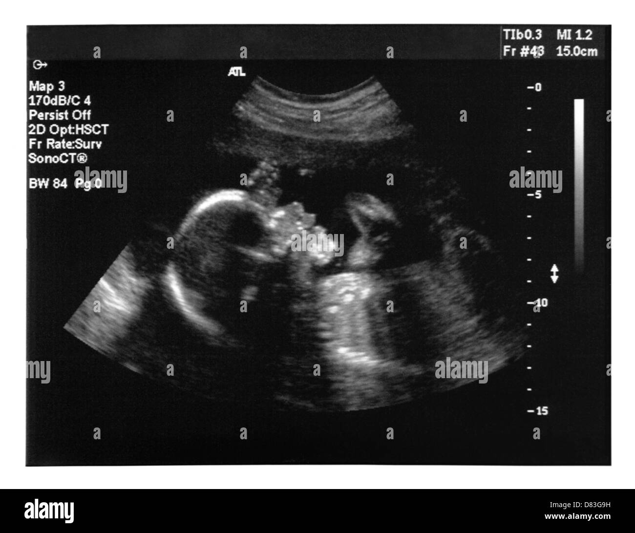
BLUE ULTRASOUND SCAN OF A 2 MONTHS OLD FOETUS Stock Photo – Alamy
i’m going to make it (after all): Ultrasound Check (16 Weeks)
Pin on NICU
pancreas anatomy ultrasound – Google Search | Ultrasound sonography …
Pin on Senior Project. Down Syndrome. Trisomy 21.
How To Get The Best Ultrasound Picture At 12 Weeks – PictureMeta
Transvaginal ultrasound of right adnexa showing the right tubal ectopic …
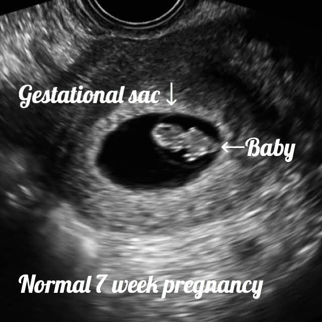
Normal 7 week baby ultrasound.
Ultrasound images of embryo or fetal development at various stages of …
Colour Doppler Ultrasound of the temporal artery before (a) and after …
Transvaginal ultrasound showing a large cervical cancer in longitudinal …
Ultrasound imaging of a baby. Five months old fetus Stock Photo – Alamy
Ultrasound image of small atrophic ovaries and hypoplastic uterus …
6 Ultrasound evaluation of an AVF. (a) Photograph showing an arm with a …
Funny ultrasound pictures
(A) Typical ultrasound image. (B) Schematic representation of …
Did You Find Out It Was Twins At Later Ultrasound? Twin Ultrasound …
Example of ultrasound measurement of subcutaneous fat thickness and …
right outflow tract ultrasound | Ultrasound technician, Medical …
The anatomy scan ultrasound is an amazing experience: you’ll get a …
Ultrasound showing stones within gallbladder and CBD References: Kfmmc …
Pelvic ultrasound showing a complex fluid collection in the pelvis …
Benign and malignant polyp. (A,B) Ultrasound depicting multiple small …
Retroperitoneal ultrasound demonstrating the pelvic mass located just …
3D Ultrasound at 30 Weeks: From Bonus Mom, to Bio Mom #3DUltrasound …
5 Week Ultrasound Pictures : 5 week ultrasound and high hcg …
3D/4D Ultrasound Pictures | Baby ultrasound pictures, 4d ultrasound …
Ultrasound of the right parotid gland region showing uneven …
Ultimate guide for your 17 weeks Ultrasound, fetal development …
Transrectal Ultrasound. | Download Scientific Diagram
(a) Transverse ultrasound of a patient with proven CTS (CTS) shows the …
Ultrasound of an AAA in the axial plane. Notes: (A) The OTO AP …
Ultrasound image of the adductor canal and puncturing needle …
Ovarian ectopic pregnancy. Transvaginal ultrasound showing a …
Ultrasound image demonstrates a hypoechoic mass, 1.9 × 0.4 × 1 cm …
Pin on superficial
Endoscopic ultrasound showing a marked dilation of the pancreatic main …
Ultrasound appearance of infected pancreatic necrosis before and after …
Medical ultrasound, Ultrasound sonography, Ultrasound school
Fake Ultrasound Pictures Fake sonogram Prank Ultrasound | Etsy
Transabdominal ultrasound of the uterus | Download Scientific Diagram
ultrasound of right ventricular overflow of fetal heart | Ultrasound …
Ultrasound image of the liver, showing multiple hyperechoic calcifying …
Normal breast ultrasound | Breast | Ultrasound sonography, Thyroid …
Ultrasound of the thyroid (transverse) showing a hyperechoic nodule …
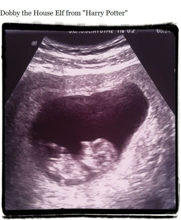
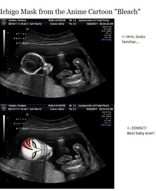
Uncanny Ultrasound Pictures (14 pics) – Izismile.com
Selected chest ultrasound images. a) Sea-shore sign. Granular …
Ultrasound of epidermoid cyst on the sole of the foot. The ultrasound …
Ultrasonographic images of different stages of corpus luteum …
Transvaginal pelvic ultrasound. Intrauterine gestational sac with Yolk …
Uncanny Ultrasound Pictures (14 pics) – Izismile.com
We extend our gratitude for your readership of the article about
how to make copies of ultrasound pictures at
galleryz.online . We encourage you to leave your feedback, and there’s a treasure trove of related articles waiting for you below. We hope they will be of interest and provide valuable information for you.


