Collection showcases captivating images of how to read a bone scan image galleryz.online
how to read a bone scan image
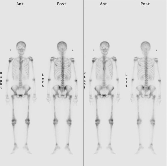
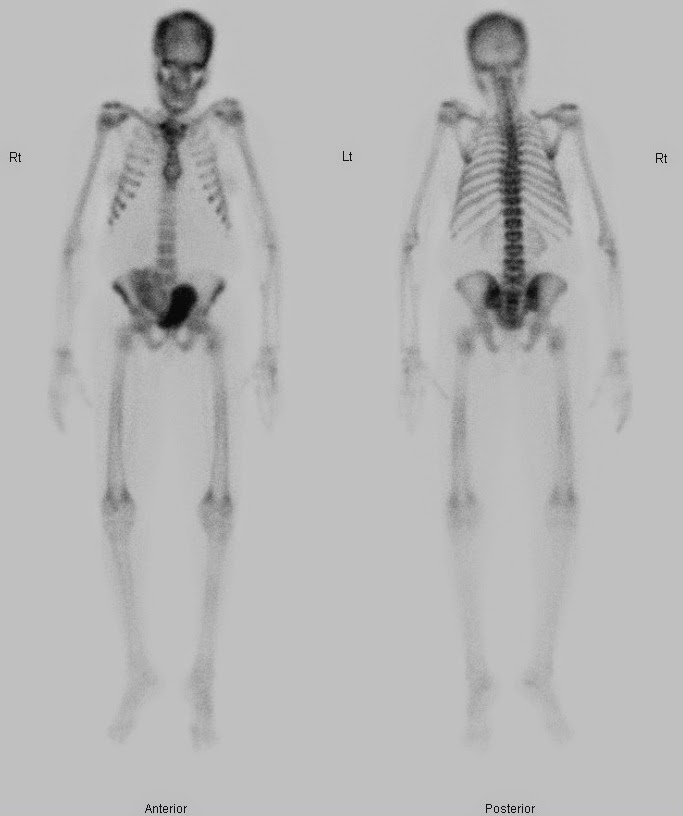
My Bone Scan test, full body by oldbogus on DeviantArt
ilaxSTUDIO » I don’t know how to read a bone scan
A bone scan is a nuclear medicine (scintigraphic) study that makes use …
Identifying Bone Metastases | Radiology Key
Image | Radiopaedia.org
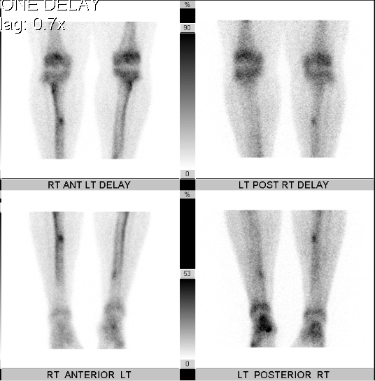
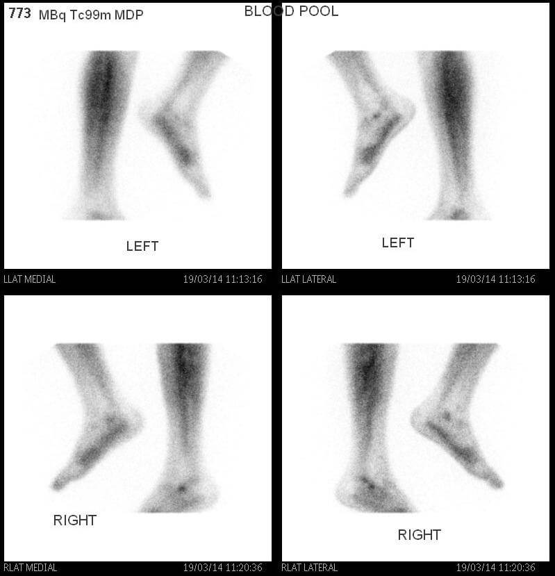
3-phase Tc99m MDP bone scan R/O osteomyelitis of R great toe : Radiology
Pre-treatment bone scan (left) showing metastases in left ribs. Bone …
Radionuclide bone scan to diagnose or confirm Avascular Necrosis …
RACGP – Bone scans
(A) Whole body bone scan reveals multiplehot spots at anterior and …
NuCleaR MuNkeE: Bone Scans and CPR
Schematic bone scan classification system. | Download Scientific Diagram
UCSD Musculoskeletal Radiology
Help me do my essay bone density scan – thedrudgereort566.web.fc2.com
-Whole body radionuclide bone scan showing high tracer uptake at the …
Nuclear Medicine & PET: Bone Scan
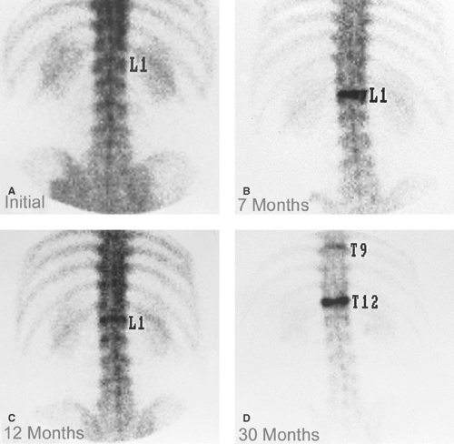
Bone scans in the last 4 years. Image C corresponds to the bone scan at …
Services – Sheila Kar , MD, FACP, FACC
Whole body nuclear medicine bone scan showing increased uptake in the …
Bone Scan | Indulge in the fascinating world of
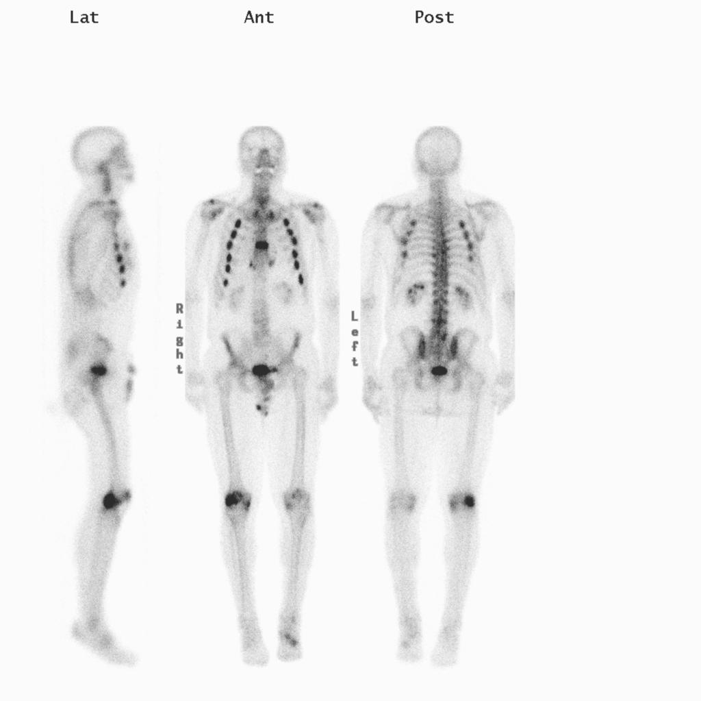
A: Anterior and B: posterior views of a whole-body bone scan performed …
Bone scan in March 2015. New progression with new lesions at the 8th to …
Sample images of successive whole-body bone scans of a patient …
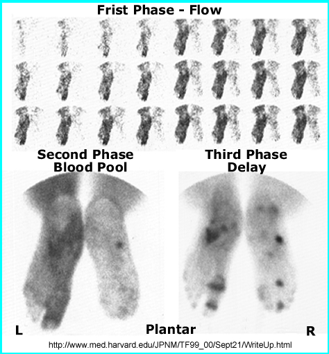
The three phase bone scan shows no specific abnormal finding at the …
Three-phase bone scintigraphy revealed mild hyperremia to the left leg …
Bone scan of the patient. Abnormally increased uptake of radioisotope …
Three phase bone scan findings of the poststroke complex regional pain …
UCSD Musculoskeletal Radiology
The Role of 18F-Sodium Fluoride PET/CT Bone Scans in the Diagnosis of …
Services – Sheila Kar , MD, FACP, FACC
Whole-body bone scan demonstrating metabolic bone disease | Download …
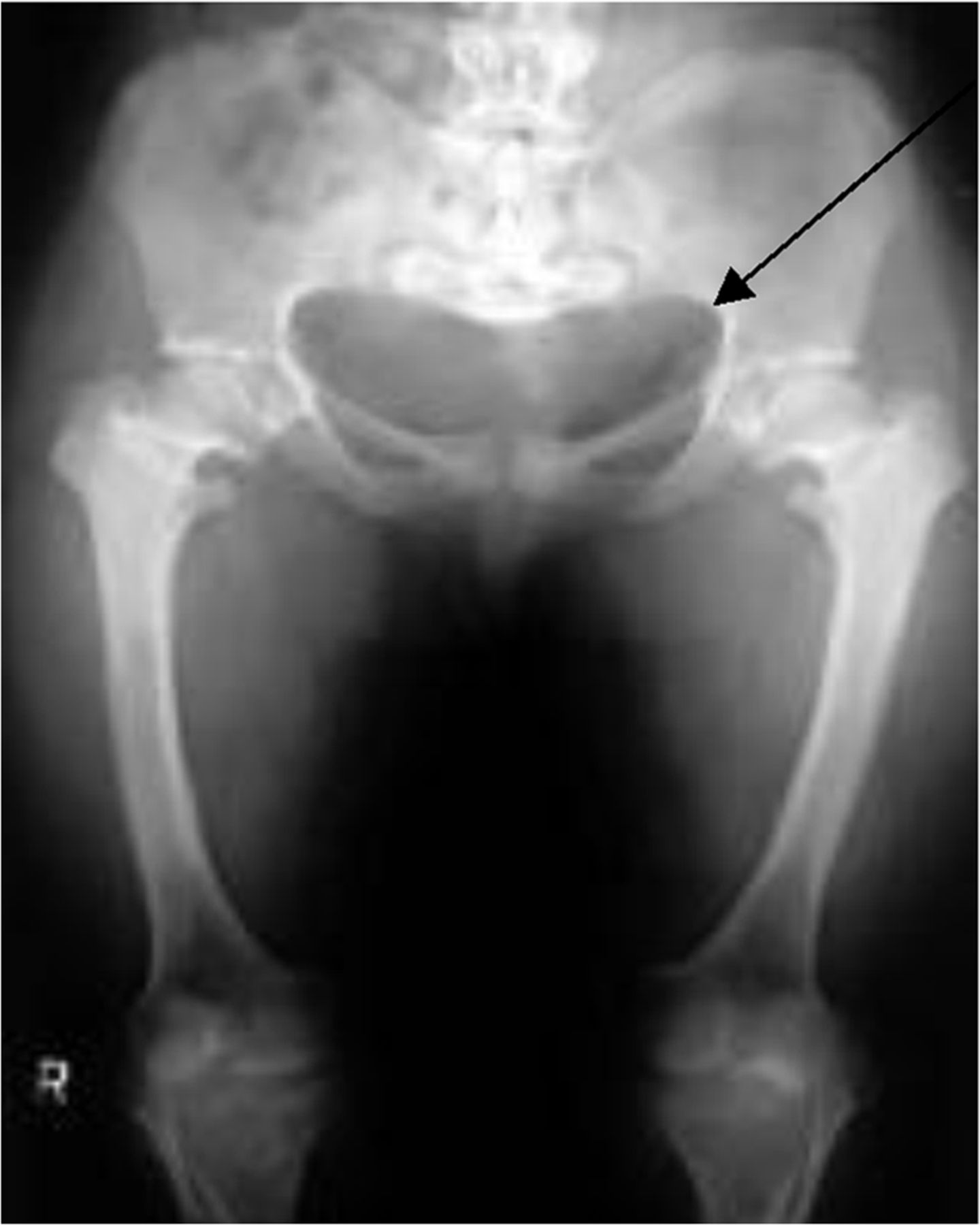
A whole body bone scan reveals that uptake of the left sacroiliac joint …
Triple-phase bone scan and white blood cell labeled scans with …
Figure 1 from Diffuse increased uptake on bone scan: super scan …
(A) Anterior amd (B) posterior radionuclide bone scans showing an …
Bone Scintigraphy – الدكتور ابراهيم عبد الرزاق البطيحي
Skeletal System Scintigraphy | Radiology Key
HPOA demonstrated on Bone Scan with increased peripheral periosteal and …
Increased Sacral Uptake on a Bone Scan with SPECT/CT in a Patient with …
hip bone anatomy
Sample images of whole-body bone scans: a previous image, b current …
Spondylodiscitis
Bone SPECT/CT imaging of the painful hip. Illustration of bone SPECT/CT …
-Nuclear medicine scan prior to diagnosis. Whole body bone scan …
An example of four-phase bone scan of a patient who subsequently …
Bone scintigraphy in a patient with active rheumatoid arthritis …
Bone scan indicated increased radiotracer uptake in several lower …
bone scans of metastatic breast cancer for 60 years old female using …
The Comparison of Bone Scan and MRI in Osteoporotic Compression …
Musculoskeletal Case 3 | NucsRadiology.com
Sample images of successive whole-body bone scans of a patient …
Incidental findings in traditional nuclear medicine practice
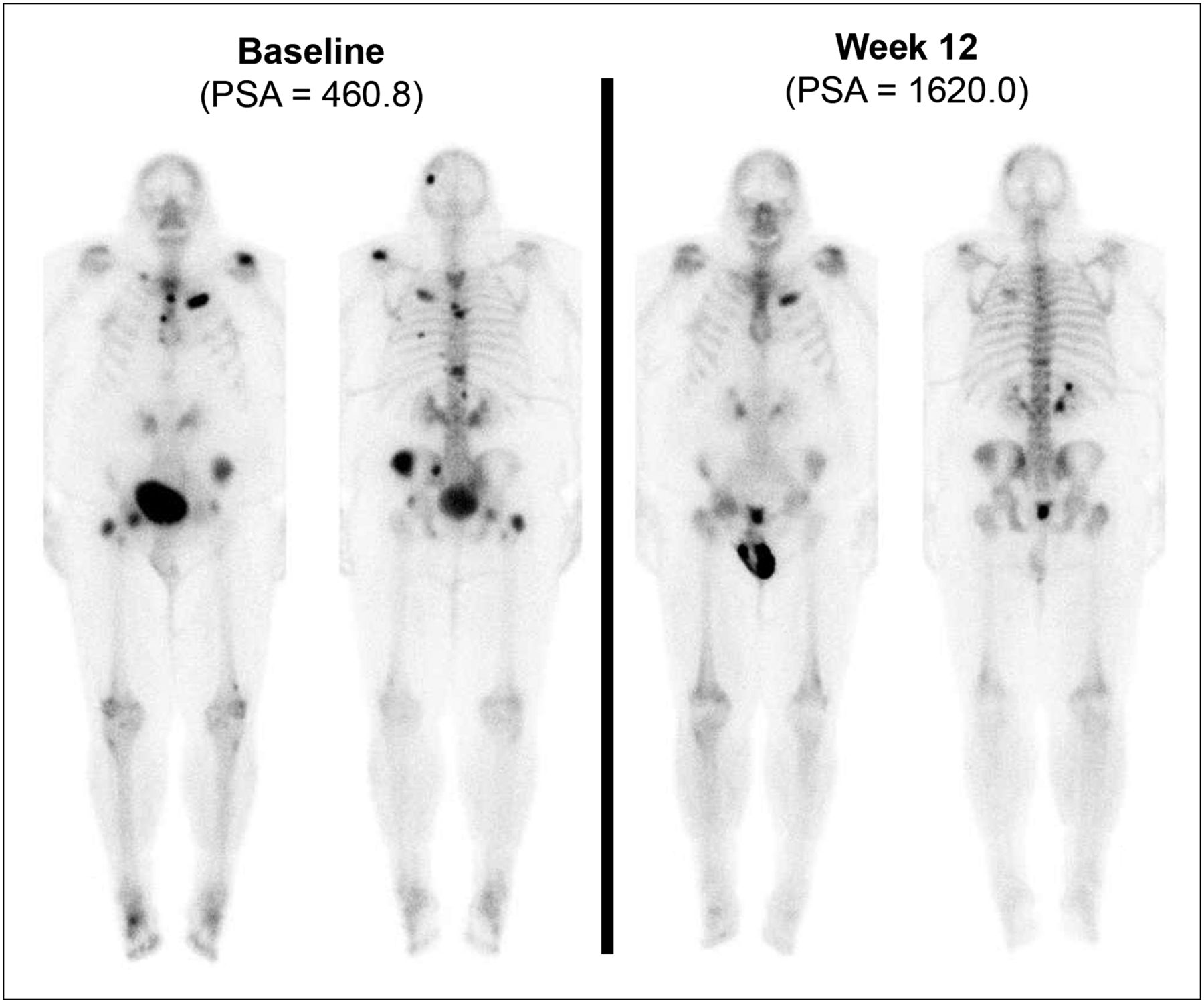
99m Tc MDP bone scans from Patient 1 (anterior and posterior) at two …
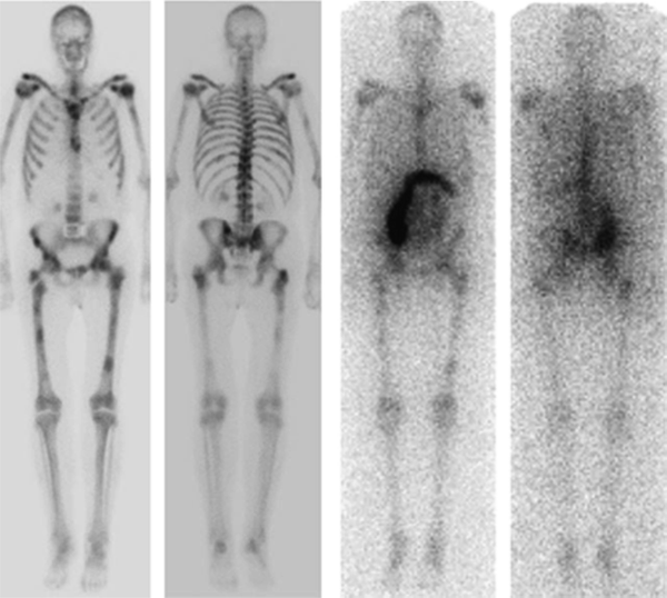
Whole-body bone scan of obese patient illustrates effect of body build …
The baseline whole body bone scan. Abnormal increased uptake was …
Management | The Bone School
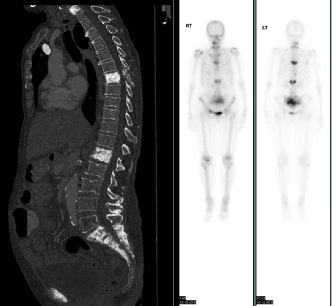
The cervical spine CT scan showed osteolytic changes with bony …
10 Nuclear Imaging | Radiology Key
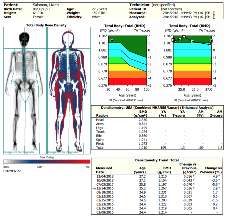
How to Read DEXA Scan Results
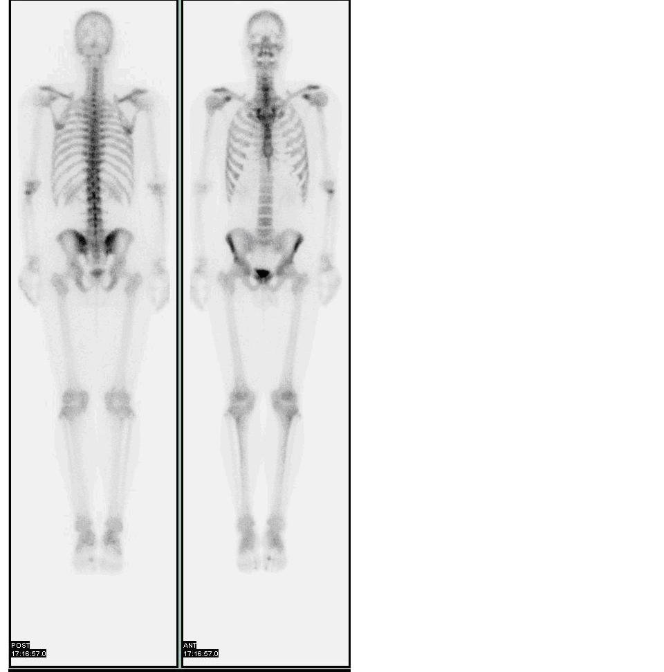
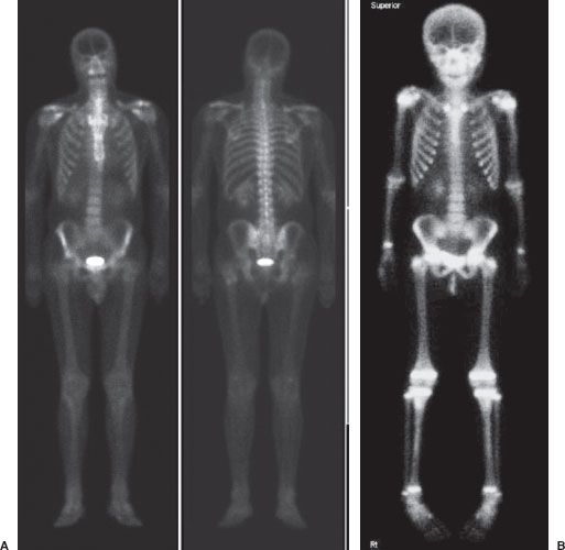
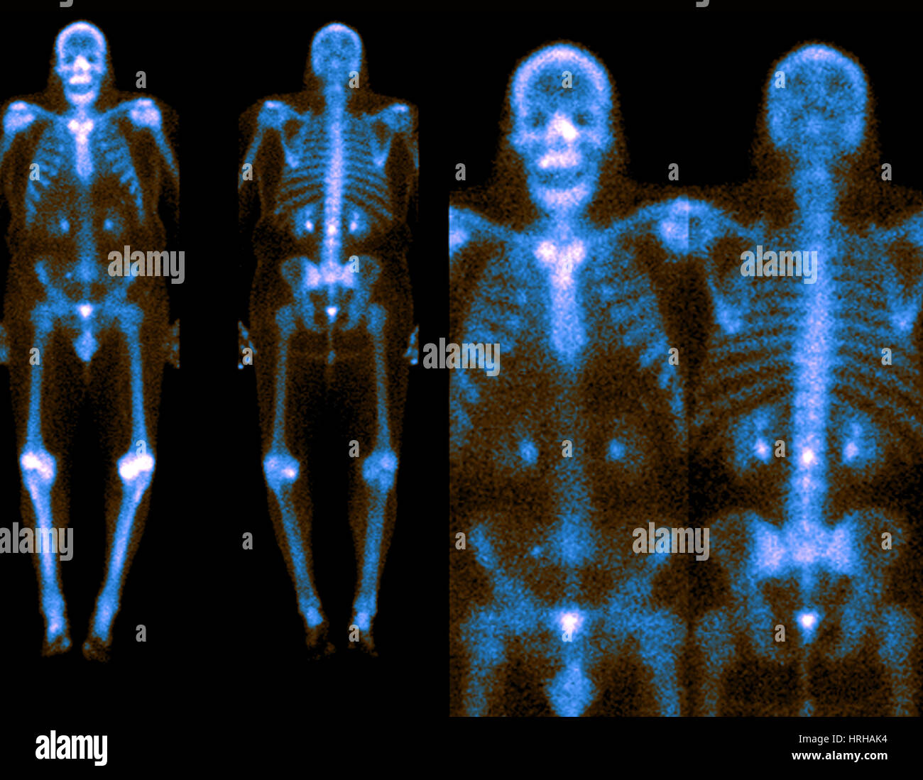
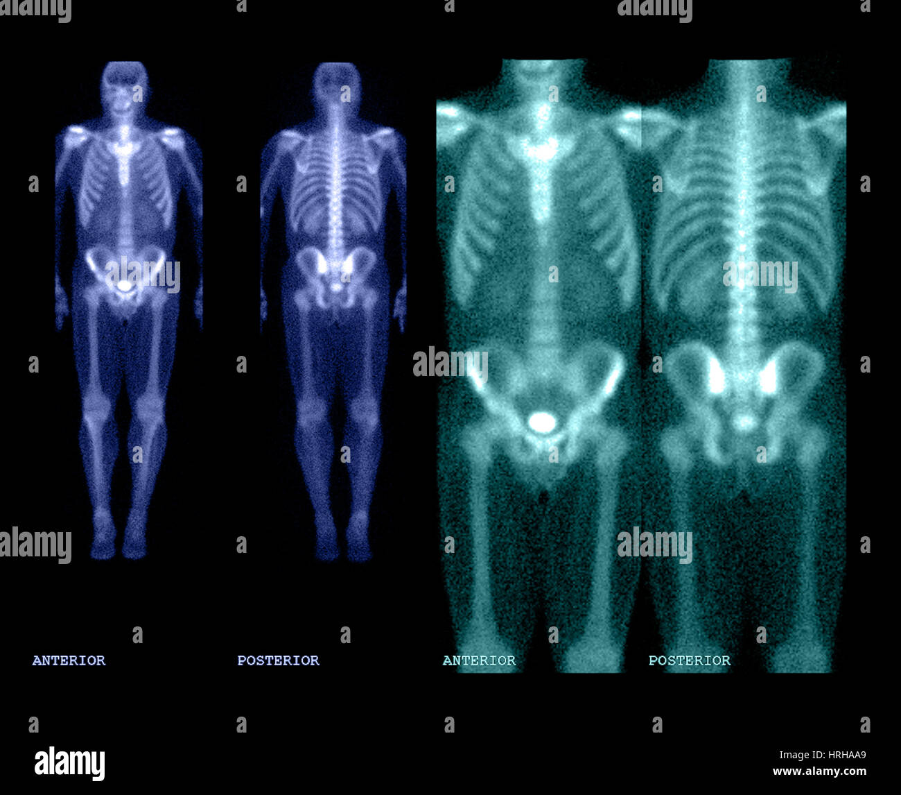
Nuclear Bone Scan High Resolution Stock Photography and Images – Alamy
Anatomy of the Bones
the xray doctor: An unusual bone scan
Bone scan (a) done before surgery, anterior planar image shows …
Injury Series: Tibial stress fractures and stress reactions: The role …
Bone SPECT/CT imaging of the painful foot/ankle. Illustration of bone …
(PDF) Long-Term Disease-Free Survival of a Patient with Oligometastatic …
[Nuclear Medicine] Bone Scan showing Pagets Disease of the skull …
Figure 6 from Unusual aggressive breast cancer: metastatic malignant …
Painful TKR | The Bone School
Phimaimedicine: 668. Bone scan
(A) Whole body bone scan reveals multiplehot spots at anterior and …
-Bone scan SPECT 3D MIP reconstructed image demonstrating asymmetric …
Bone Scans Top: Typically 2-6 hours after intravenous administration of …
Medical Pictures Info – Bone Scan
Giant Cell Tumor of Bone: Bone Tumors
SPECT/CT scans show multiple bone metastases. | Download Scientific Diagram
Temporal Bone – CT Scan – RadTechOnDuty
Bone scans of both knees in the anterior view before (left) and after …
bone scan | Medical Pictures Info – Health Definitions Photos
99mTc-HDP bone scintigraphy (BS) finidings considered as Rheumatoid …
Isotope bone scan (July 1987) showing extensively increased uptake …
Anterior projection technetium-99 m-MDP whole-body bone scans in a …
DEXA Results: 29 / 5’5″ / 127lbs / 22.4% : xxfitness
(PDF) Bone scan patterns of patients with diffuse metastatic carcinoma …
Bone scan performed in March 2014 showing metastatic disease …
View Bone Scan Images Showing Cancer Images
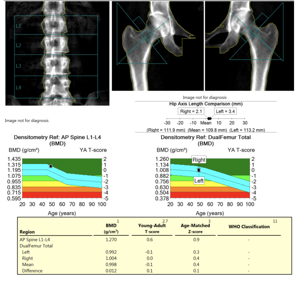
Hologic bone density report sample
Bone SPECT/CT imaging of referred pain. Illustration of bone SPECT/CT …
Tarsal Coalition | Mr Malik Orthopaedic Consultant
Nuclear Bone Scan High Resolution Stock Photography and Images – Alamy
Predicting Height by Bone Age… An Inexact Science…
(PDF) Biochemical Recurrence in Prostate Cancer and Temporal …
Historical Perspective on the Use of Radionuclides for Therapy …
introduction of anatomy
Three-phase bone scan, revealing increased uptake at the third …
Diagnostic modalities of osteoporosis. (A) DXA bone scans of the lumbar …
Bone scan (A) and preoperative MRI (B) of a grade I. chondrosarcoma in …
PET/CT scan of a patient with breast cancer. A trace amount of …
VIDEO
All about osteoporosis in older adults – Online interview
We extend our gratitude for your readership of the article about
how to read a bone scan image at
galleryz.online . We encourage you to leave your feedback, and there’s a treasure trove of related articles waiting for you below. We hope they will be of interest and provide valuable information for you.