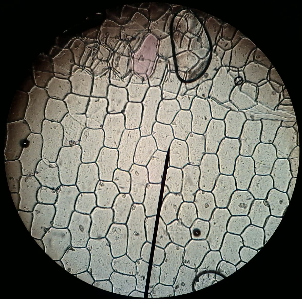
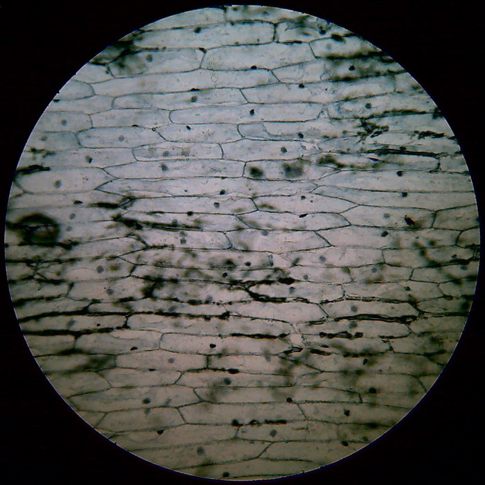
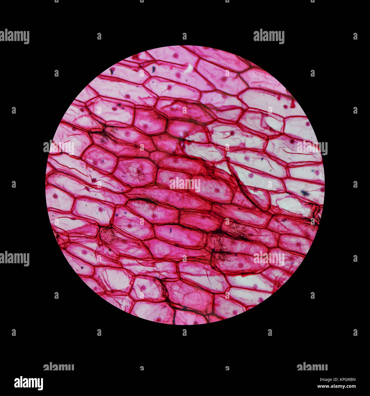
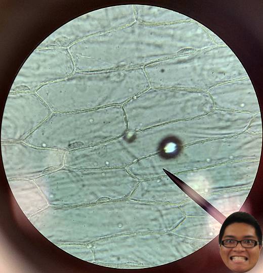
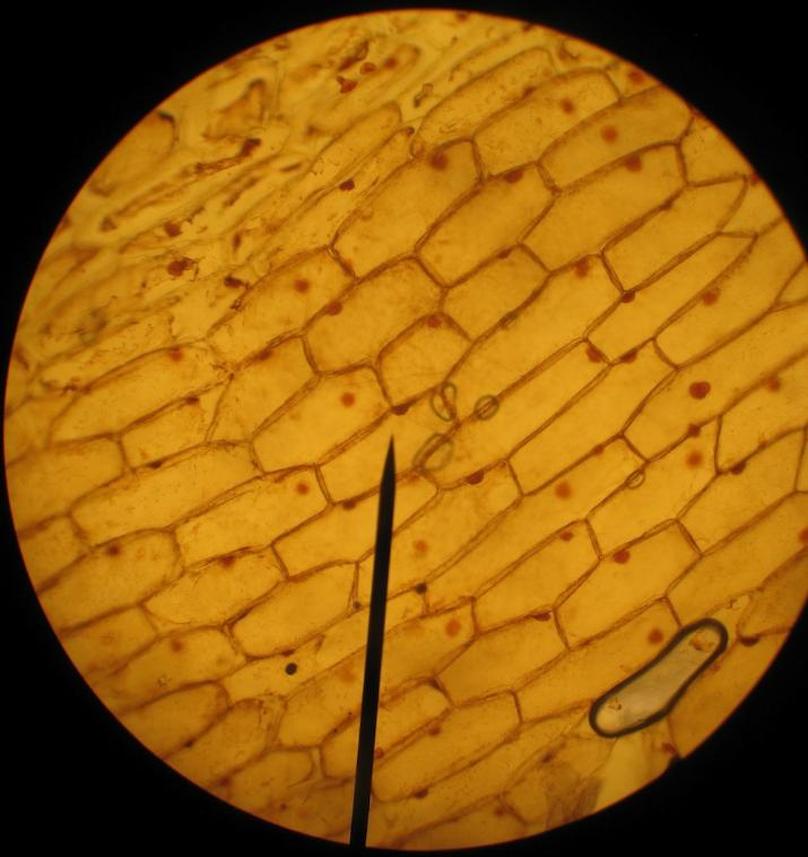
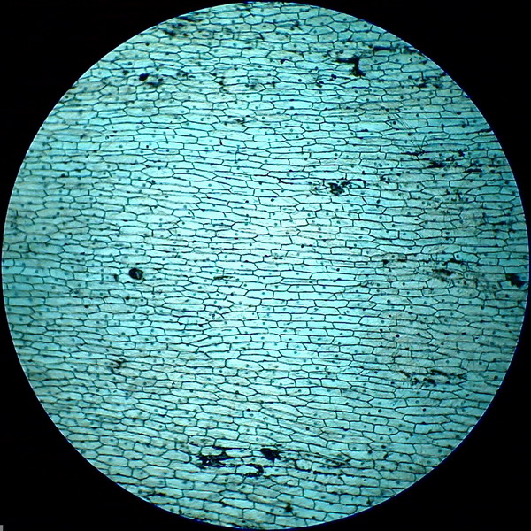
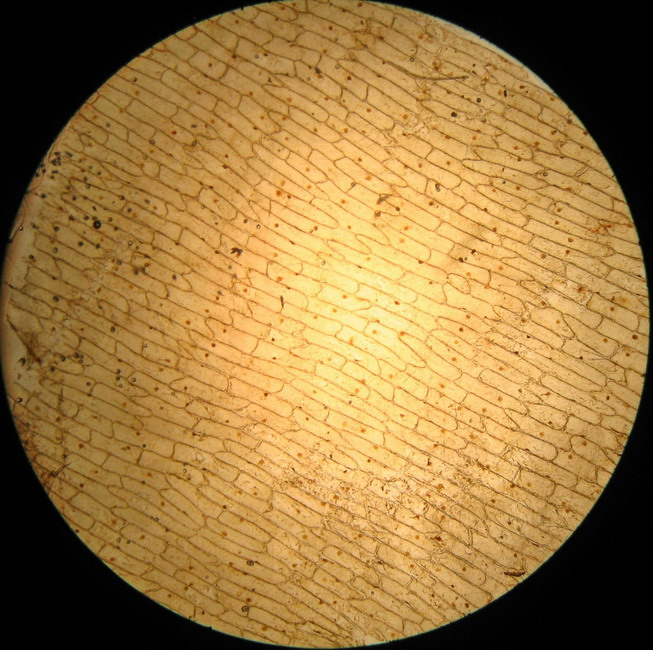
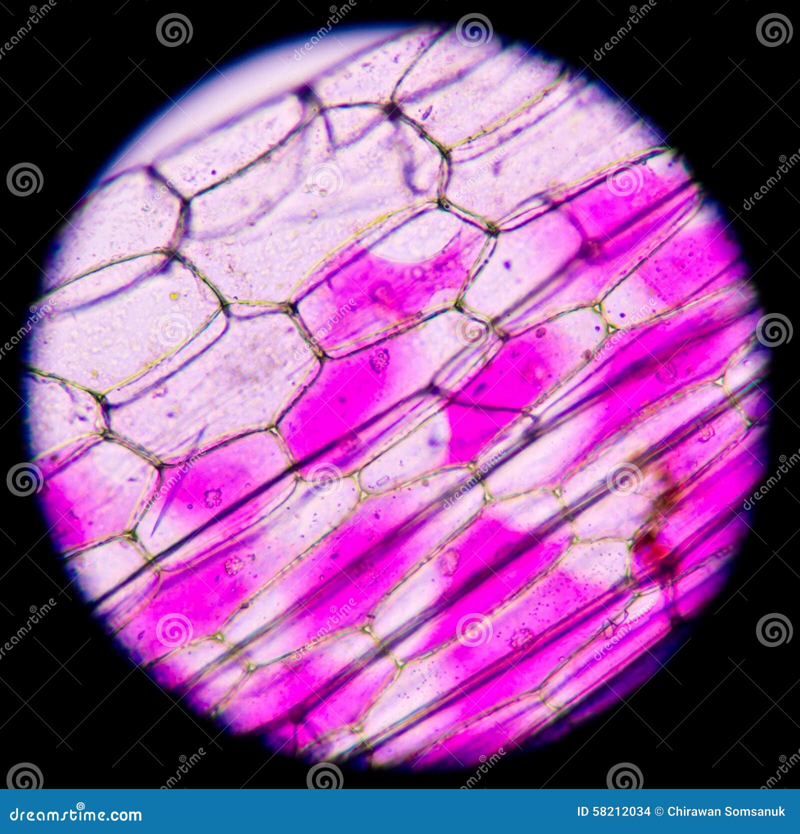
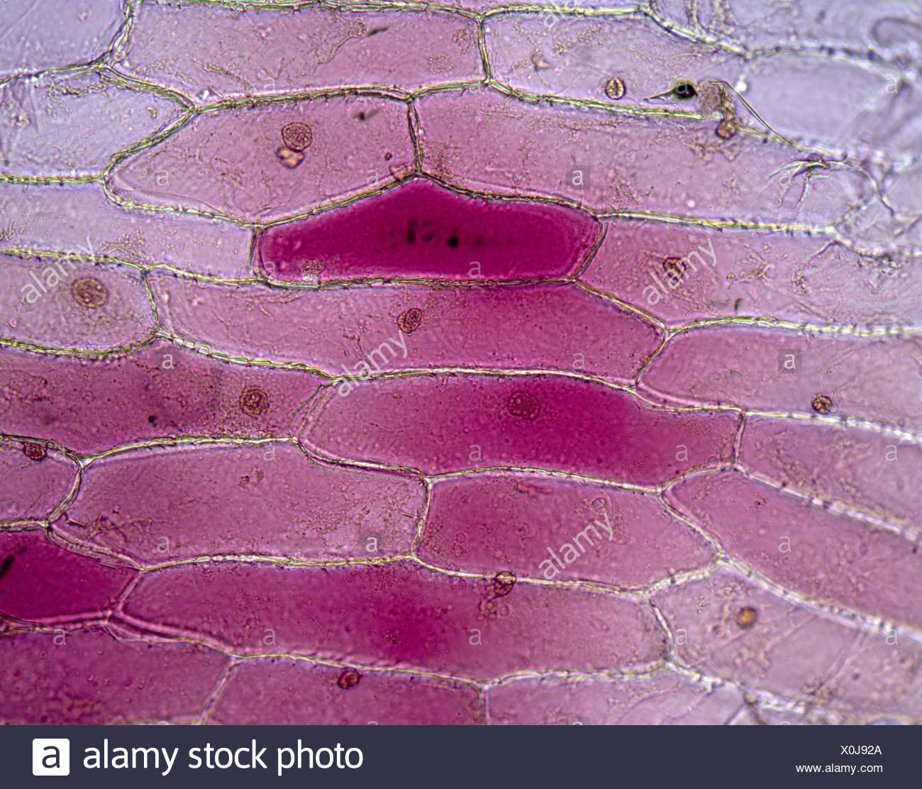
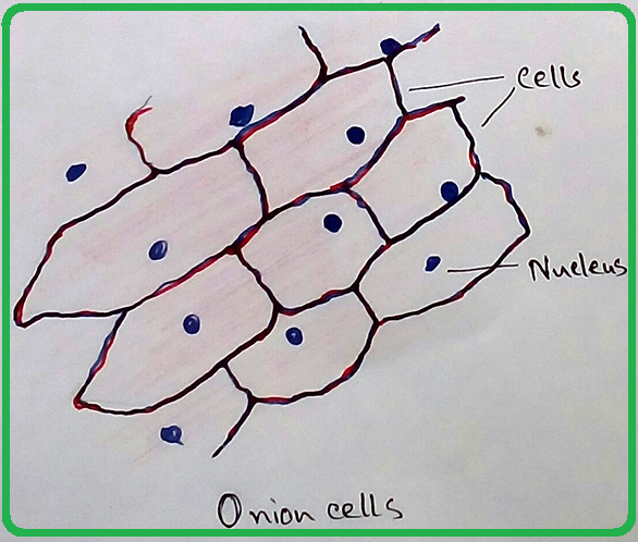
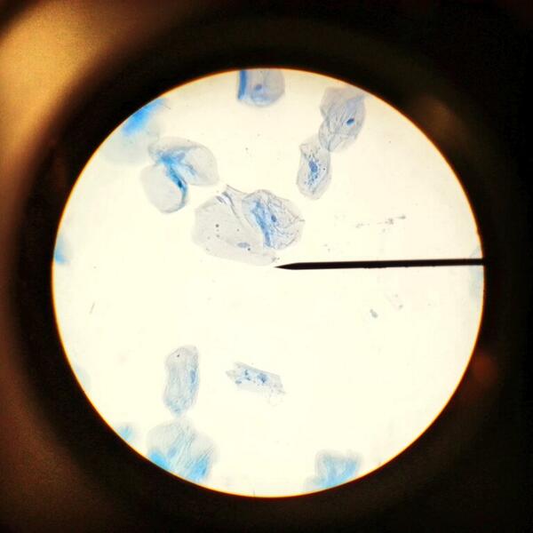
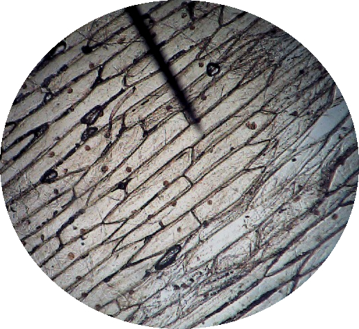
List showcases captivating images of jack is seeing an onion cell under a microscope galleryz.online
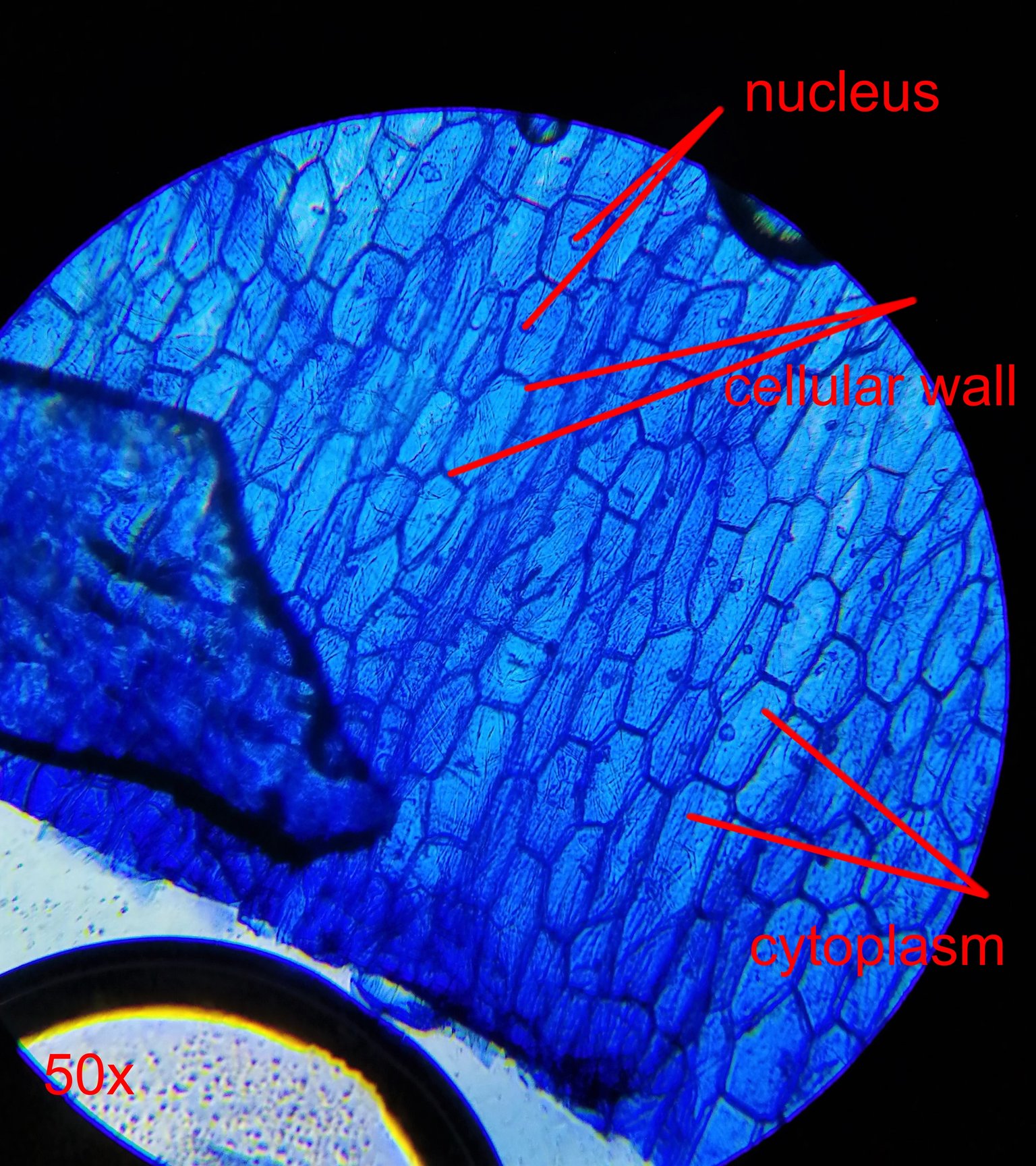
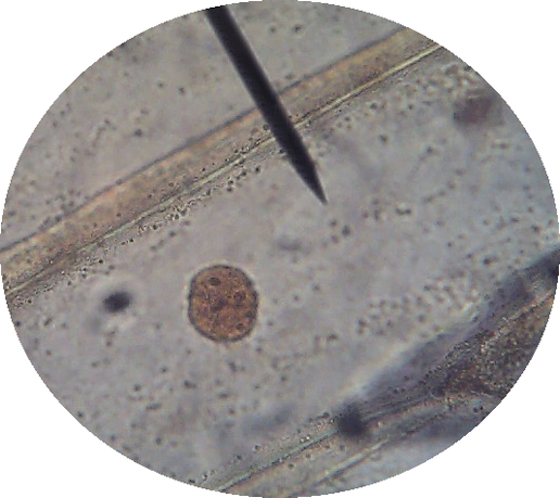
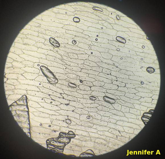
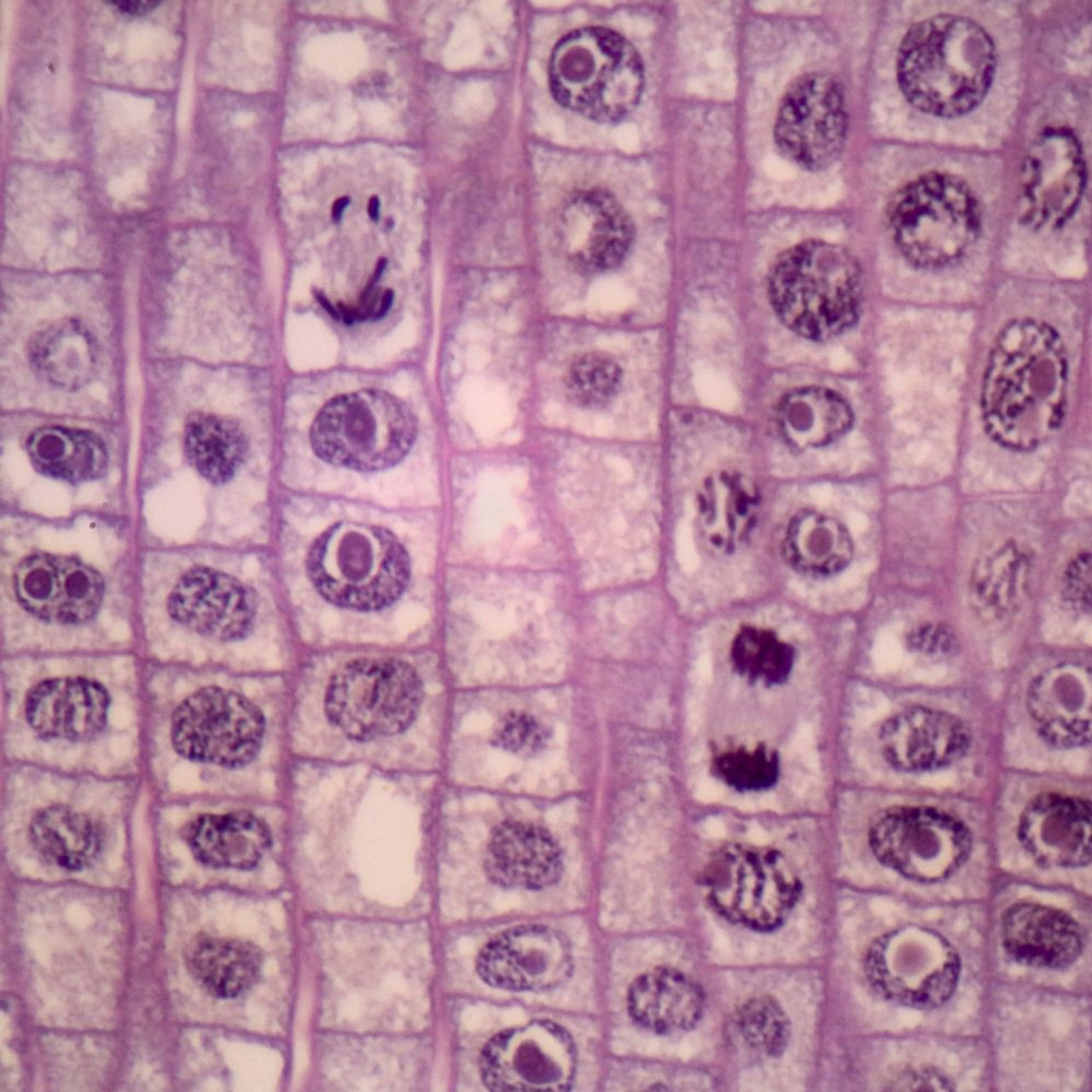
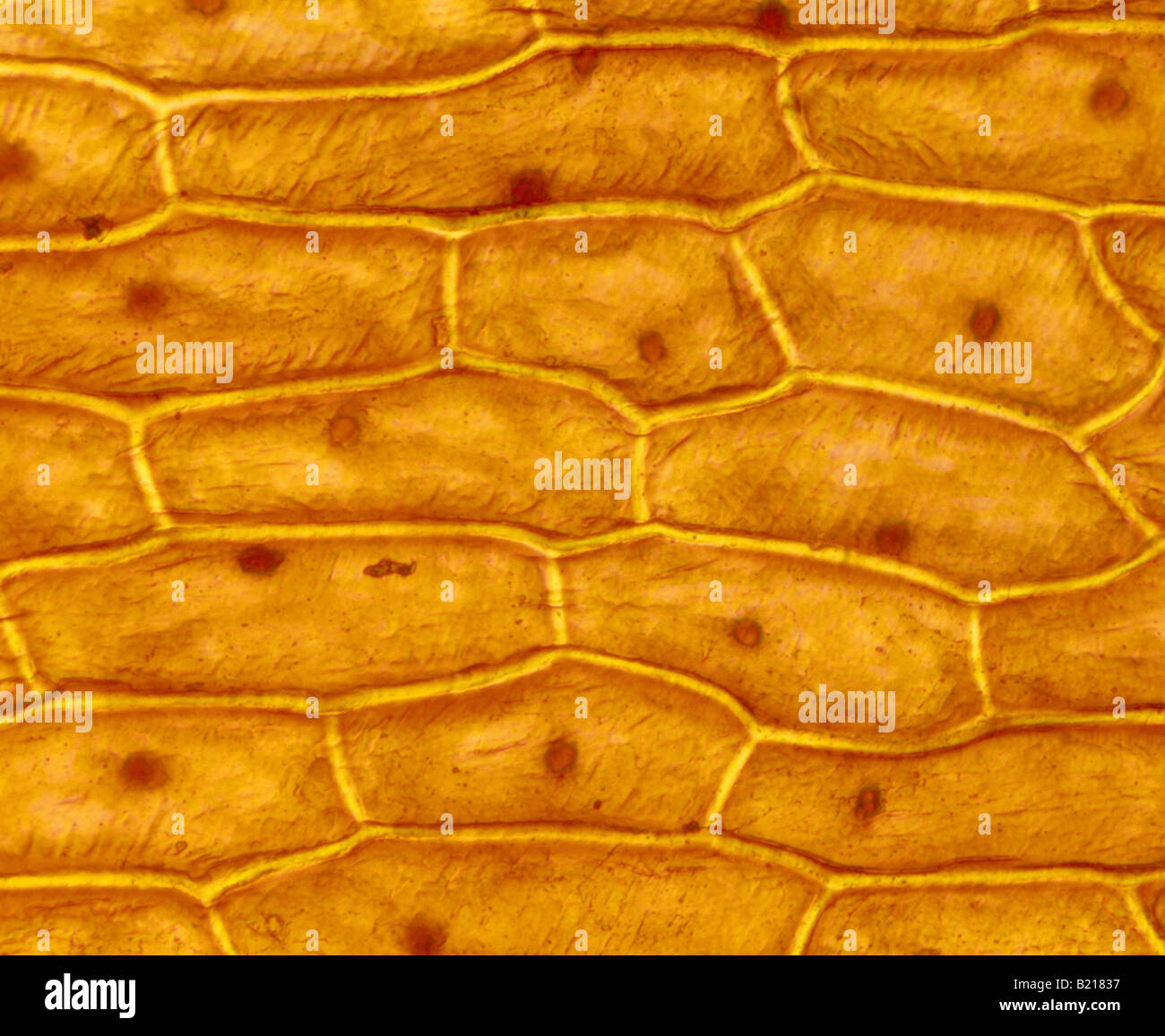
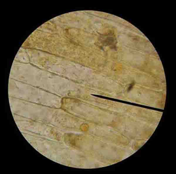
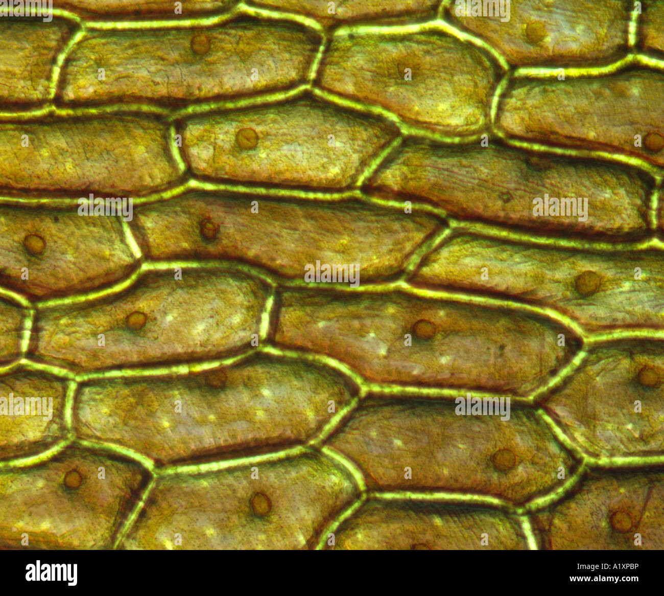
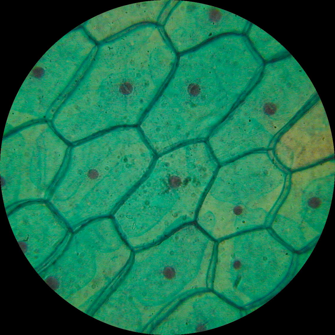
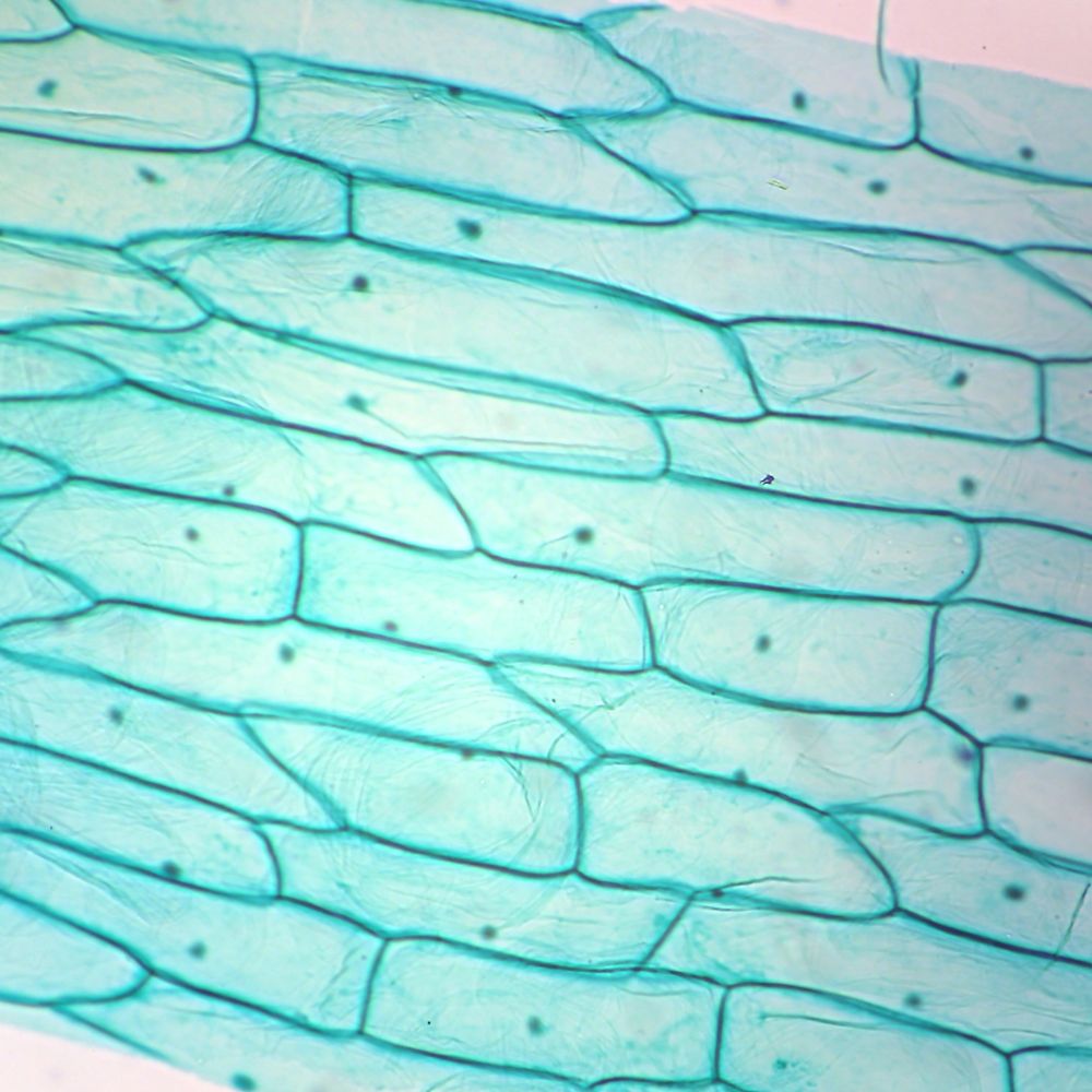
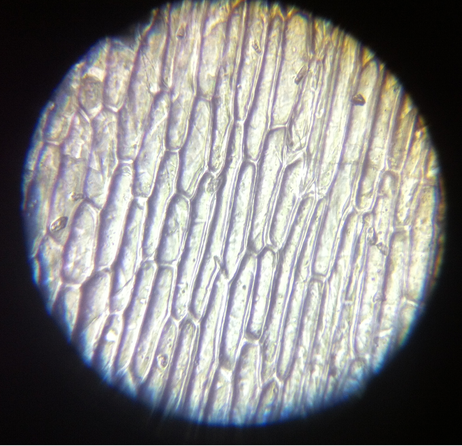
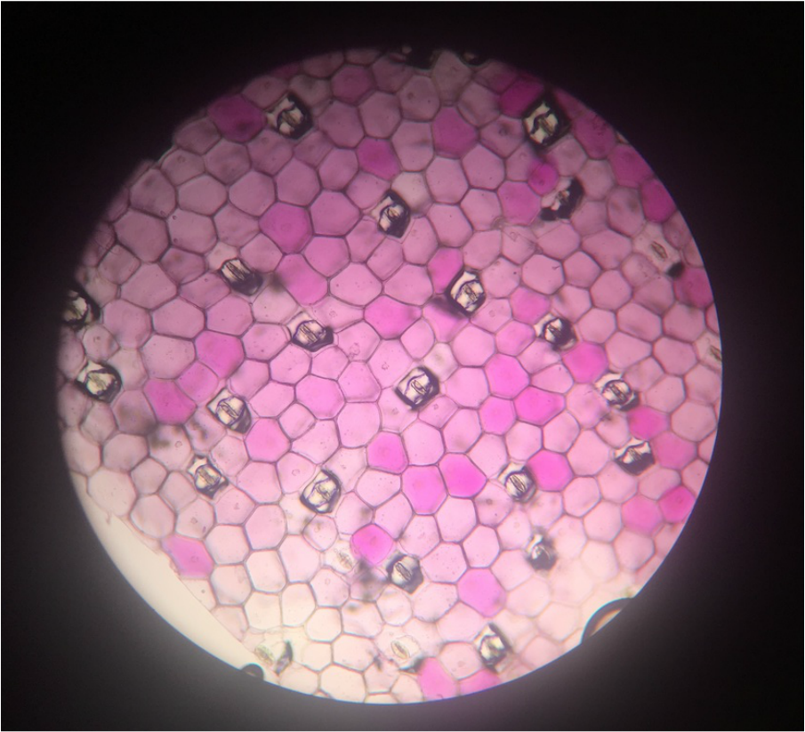
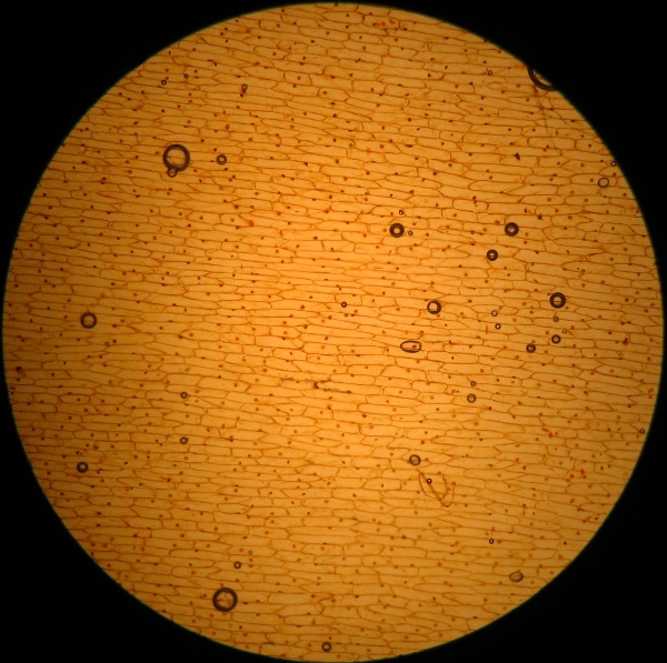
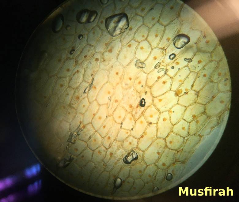
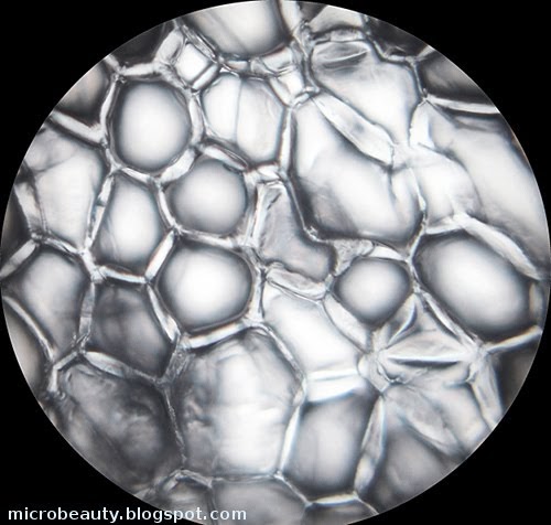
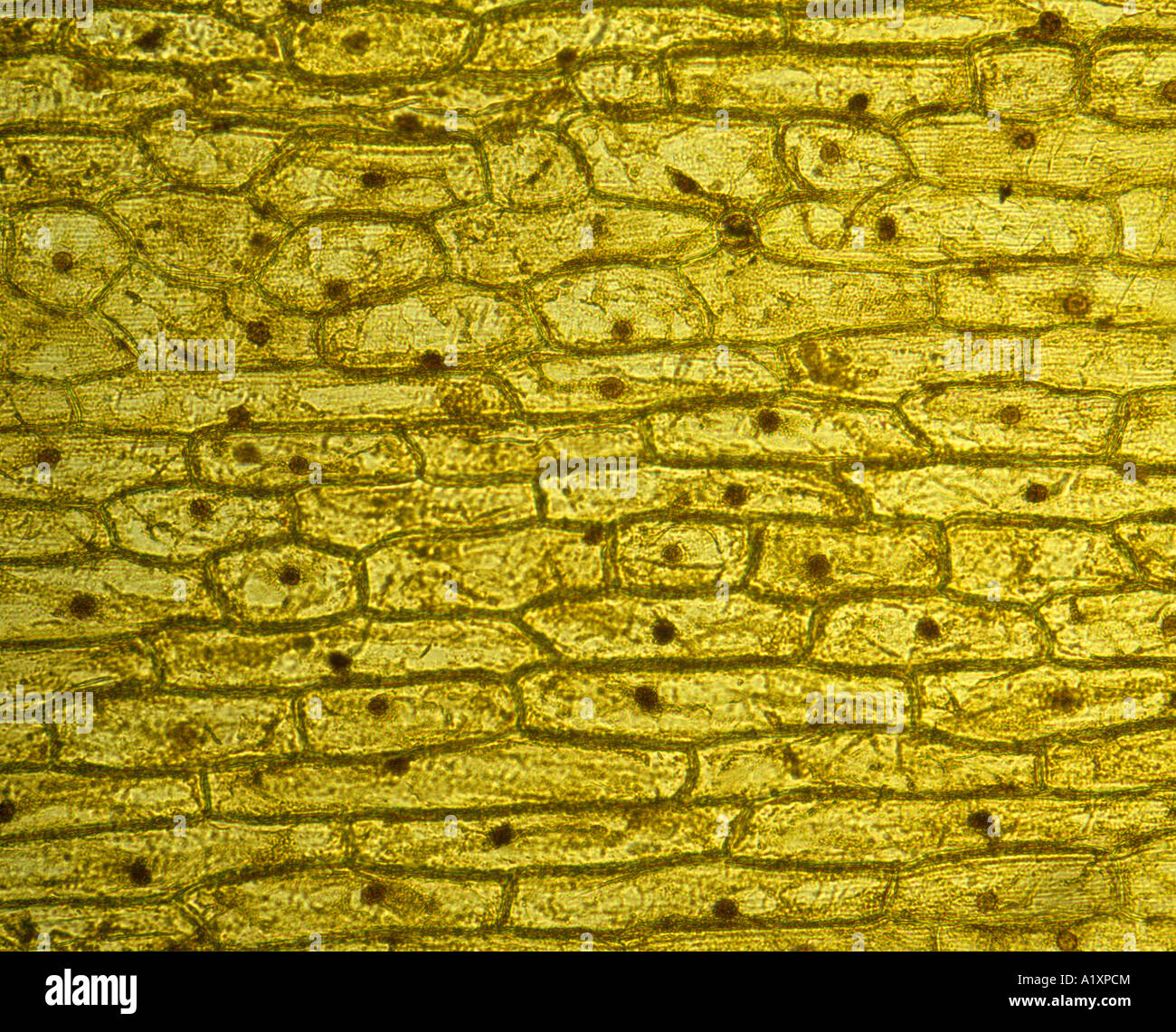
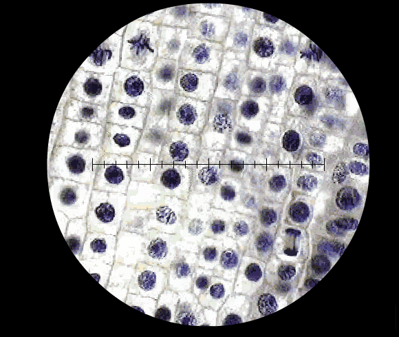
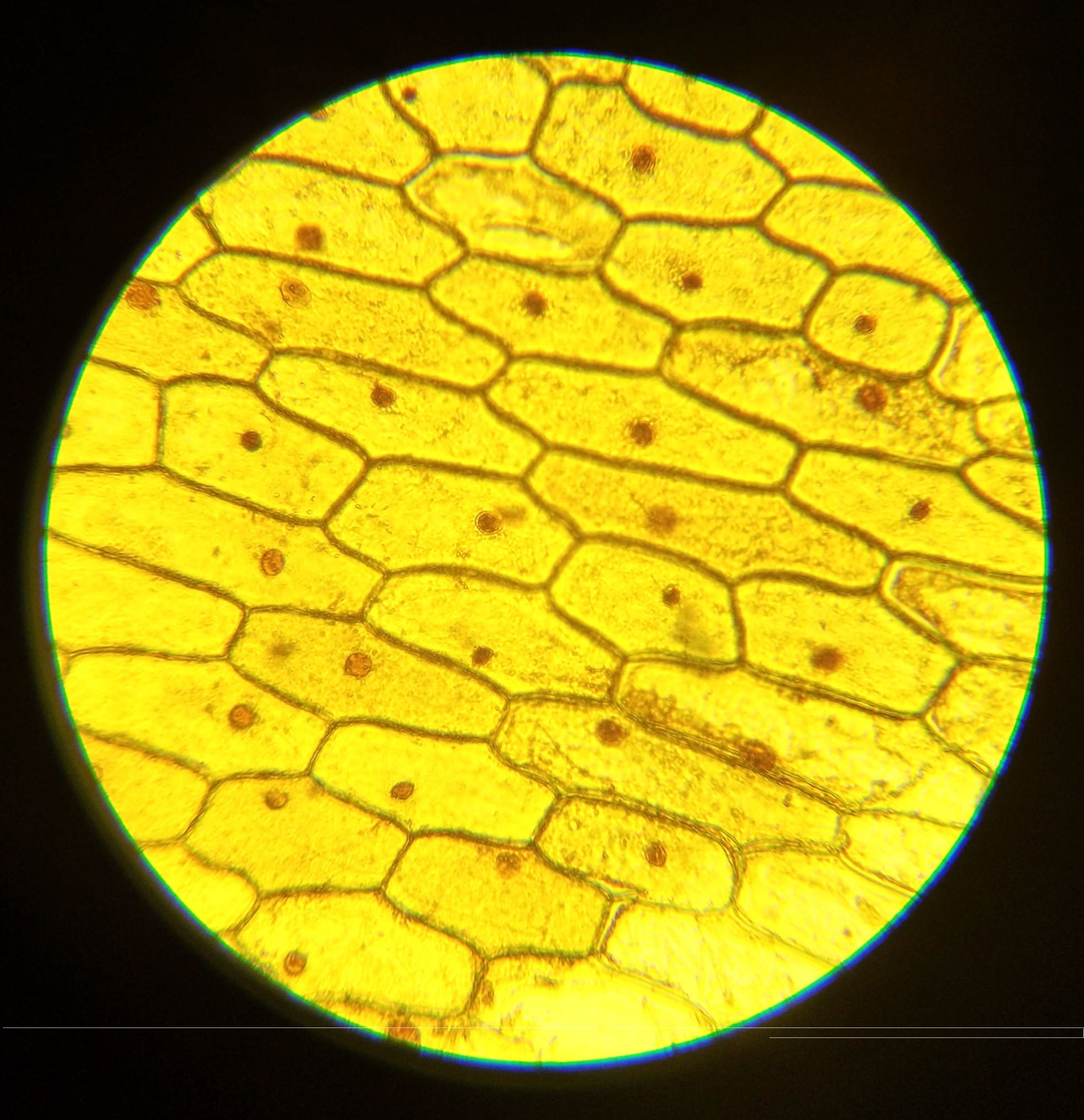
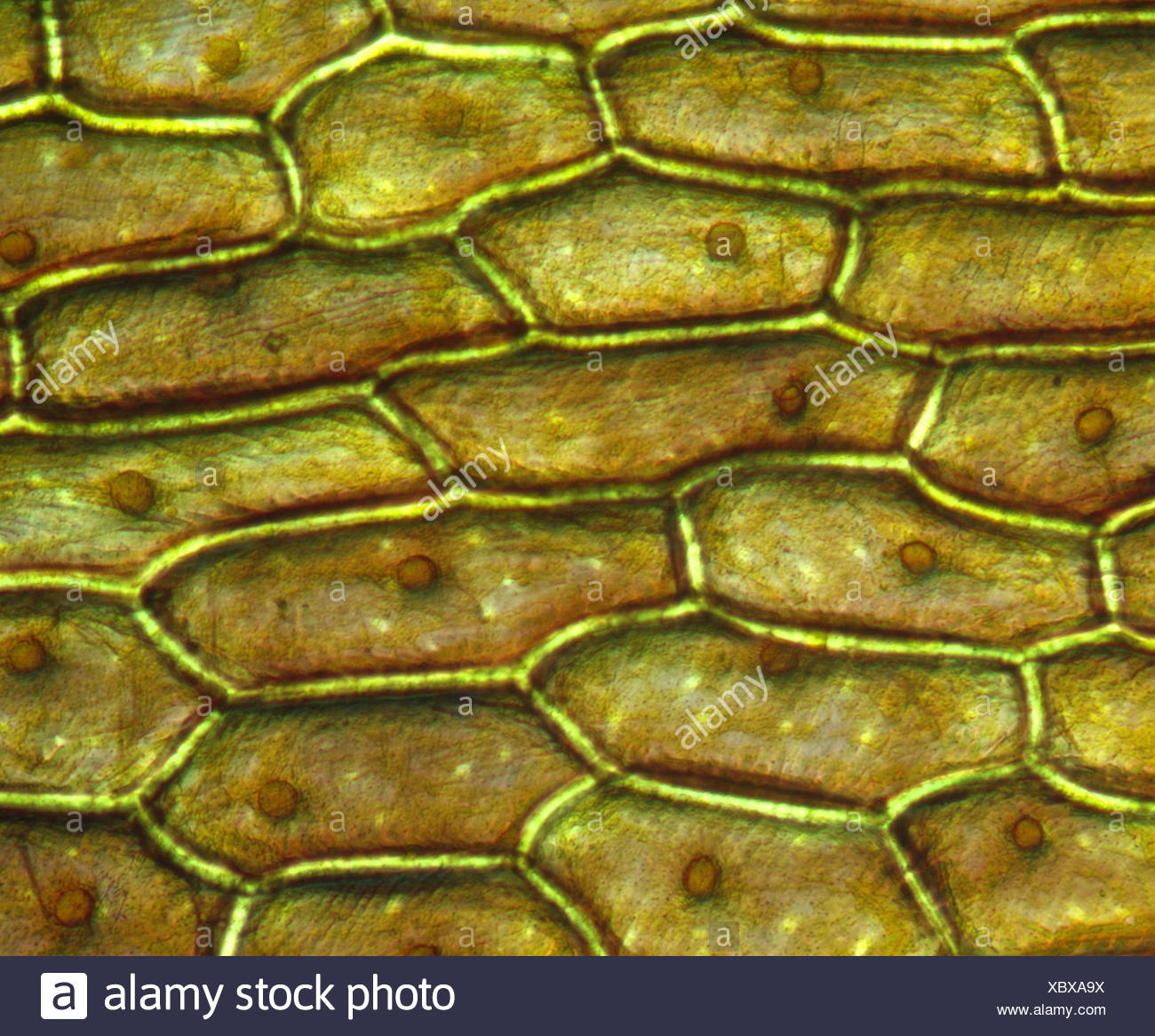
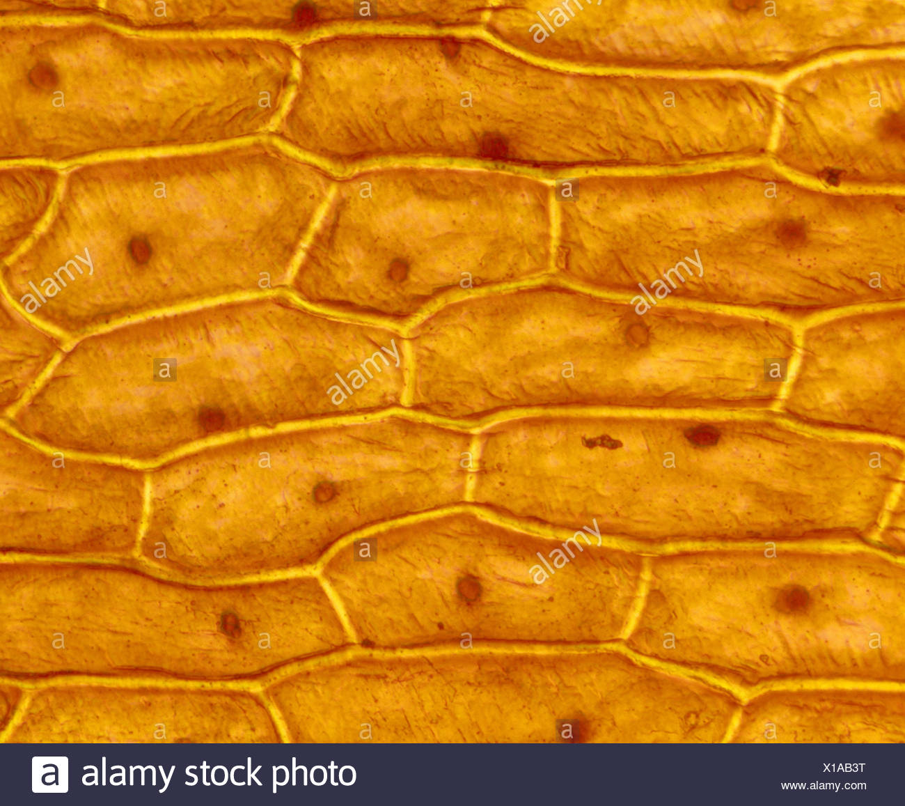
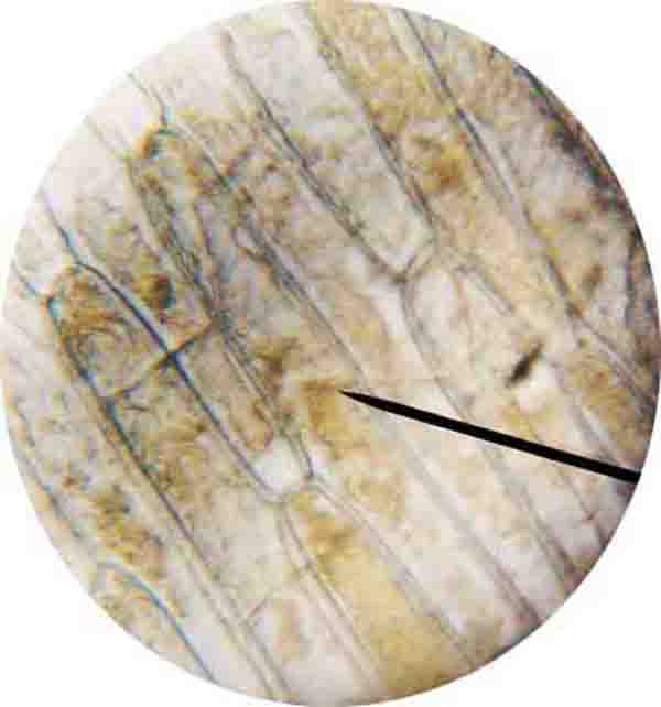
jack is seeing an onion cell under a microscope
Onion Cells | Cool science experiments, Macro and micro, Microscopic photography
onion cells Biology Art, Cell Biology, Patterns In Nature, Textures Patterns, Microscopy Art …
Grace’s Microscope — Onion cells exposed to a hypotonic solution….
Gordon Cooke » Student Images and Videos from the Microscope Practical
Title
Onion cells | High-Quality Nature Stock Photos ~ Creative Market
Onion Plant Cell Under Microscope Labeled / Onion Cells – Onion epidermis with pigmented large …
Cells key
Squash an Onion and Learn the True Age of Your Cells | Carolina.com | Science cells, Cell …
Onion Skin Cells Under Microscope submited images.
[Solved] I need the tables for both the onion cell and the cheek cell done… | Course Hero
ONION CELLS AT 400X – MicrobeHunter.com Microscopy Forum
The inner epidermis of the onion bulb cataphylls
The inner epidermis of the onion bulb cataphylls
The inner epidermis of the onion bulb cataphylls. 5) Fixing with Clarke’s fixative – Staining …
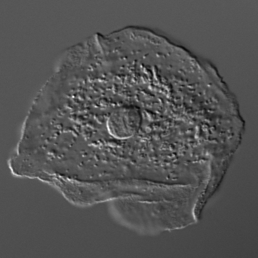
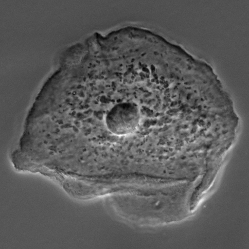
Animal Cell Under Microscope 400X : CZ29-003b.jpg | Kuhn Photo / In this experiment we will see …
DISCOVERY-IT SCIENCE (EPITHELIAL TISSUE OF ONION) OC 👨🏫👨🎓🧫🔬 — stemgeeks
My onion cells at 100x magnification! | Magnification, Celestial bodies, Cell
Plant Cell Lab (Makeup)
Cells
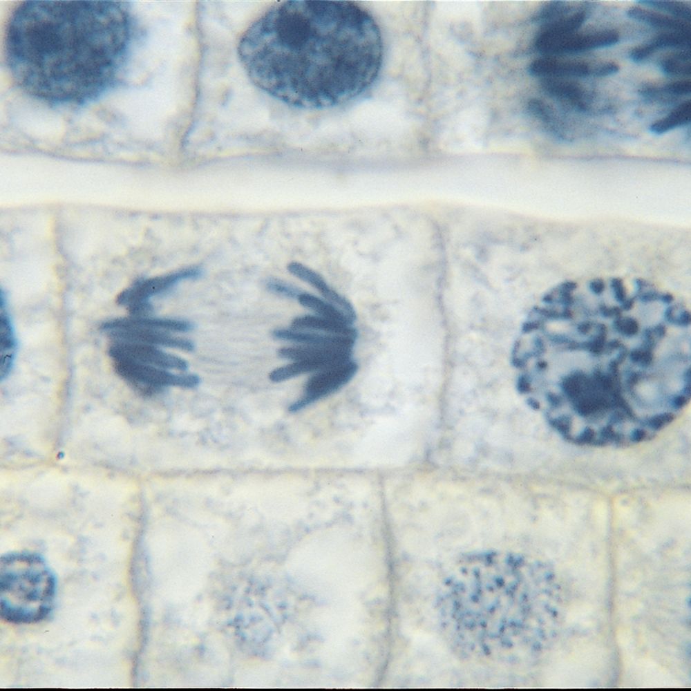
Onion Mitosis, l.s. Thin Microscope Slide – Southern Biological
Onion Plant Cell Under Microscope Labeled / Onion Cells – Onion epidermis with pigmented large …
Onion Cell Under Microscope 40X Stock Image – Image of cell, onion: 203557595
Onion cells ~ Nature Photos on Creative Market
ChaiSY’s blog: BIOLOGY NOTES FORM 4 CHAPTER 2 KSSM
The Cells and Microorganisms Webquest
Лук под микроскопом. #onion #cell #cells #onioncells #micro #microscope #biology #botany Onion …
How to Prepare a Wet Mount Slide of Eukaryotic Cells – Page 2
Microscope Onion Cell Structure – Micropedia
Chapter 8: Biology: Photography through the microscope
Plant Cell Under Microscope 400X Labeled – Untitled 1 / Why is the elodea leaf not visible in a …
Pin by Hopzie on Microscoop | Plant cell, Things under a microscope, Cell biology
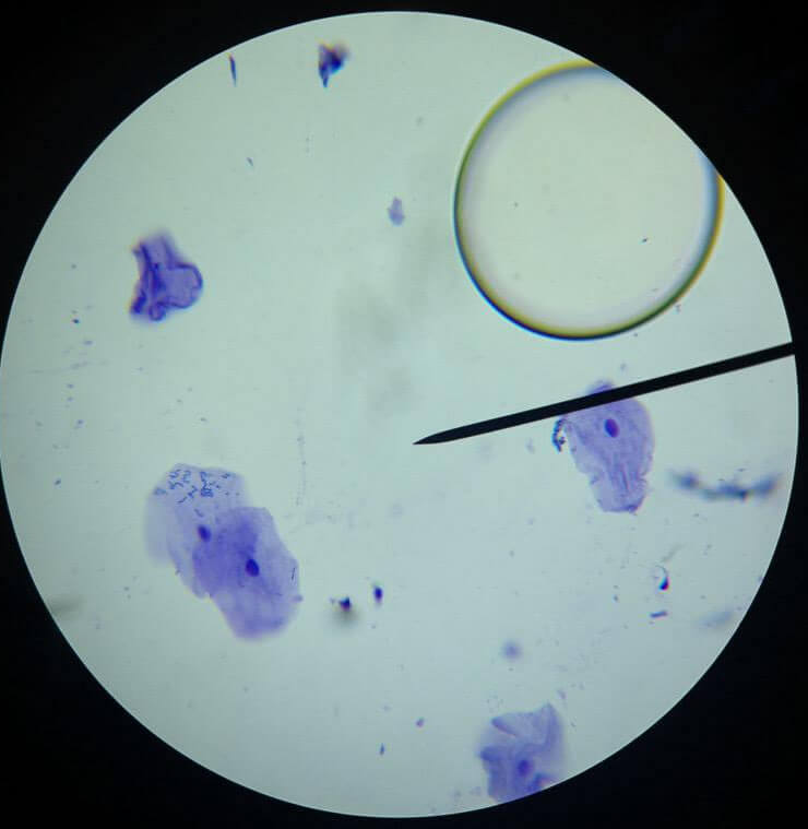
Cheek Cell Lab – Biology LibreTexts
Plant Cell Lab (Makeup)
Science Quest: Observing Onion Cells – Blog, She Wrote | Things under a microscope, Science …
IGCSE_WADI_2015-16 – BIOLOGY4IGCSE
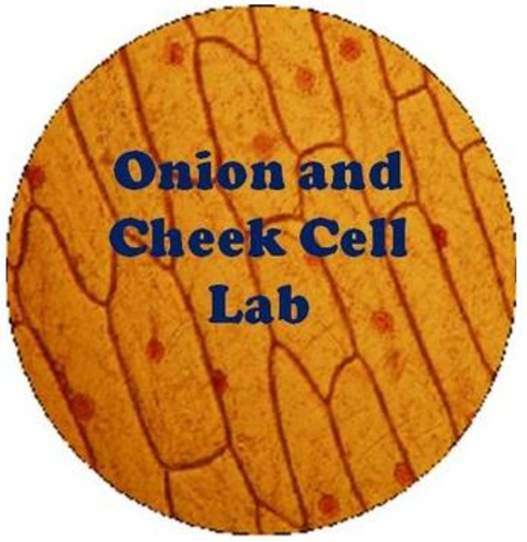
Onion and Cheek Cell Lab Experiment – Organelles | Teaching Resources
Animal Cell Microscope Slide : What does an animal cell look like under an electron …
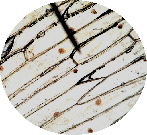
Onion Skin Under Microscope 400x | Things Under a Microscope
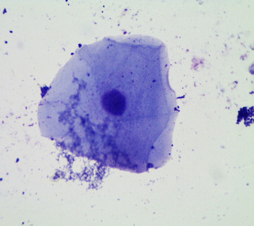
Cheek cell under microscope | Video + image – awesomeBiochem
Online Onion Root Tip Mitosis Lab – ROOTSA
Biology Photos
Slides Of Various Stages Of Mitosis Under Compound Microscope – Micropedia
onion peel cell and cheek as through Microscope diagram. – Brainly.in
Onion Mitosis, c.s., 15 µm, Hematoxylin Stain Microscope Slide | Carolina.com
Onion Root Cell Mitosis Labeled – Juventu dugtleon
Important Points of Cytoplasm – Cells – Chapter 8 Class 8 Science
Plant Cell Under Microscope 400X / Banana Plant Cell Zoomed 400x Magnification By Crixans On …
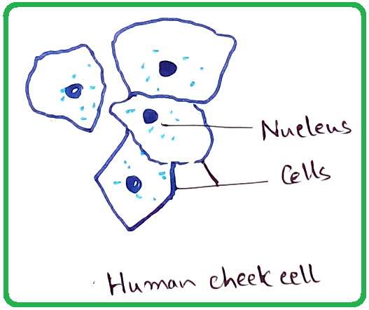
Human Cheek Cell (methylene blue stained wet mount) | Apologia biology, Prokaryotic cell …
The inner epidermis of the onion bulb cataphylls.
Animal Cheek Cell Experiment – Lab Human Cheek Cell – Onion and cheek cells were observed under …
Stained onion root tip under microscope (400X) Biosensing assay: The… | Download Scientific …
Cells under a microscope : Biological Science Picture Directory – Pulpbits.net
Cells key
👍 Red onion cell in salt water. Observing osmosis, plasmolysis and turgor in plant cells. 2019-02-07
cell-cheek-03 | Cheek Cells | biologycorner | Flickr
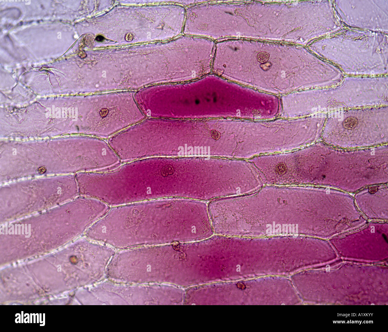
Onion Cells High Resolution Stock Photography and Images – Alamy
My Cheek Cells 7th Grade Science | Free Nude Porn Photos
Onion Peels Observed Under the Microscope | Confirmation Point
Onion_cells_under_the_fluorescence_.8 wiki | The Common Vein
Cheek cell under microscope | Video + image – awesomeBiochem
Cheek Cell Under Microscope : Cheek Cells Under Microscope – YouTube : Experiment conducted in …
The Wonderful Microworld: Onion Skin Cells (Onion Scales) (Outer most dry layer of the onion Bulb)
Transport in Plants
Plant Cell Under Microscope
Plant Cell Lab (Makeup)
Onion Skin Under Microscope | Things Under a Microscope
The Wonderful Microworld: Cell Nucleus – Onion
The Wonderful Microworld: Onion Cells – (Onion Bulb)
ONION SKIN CELLS / EPIDERMAL CELLS / STAINED IN IODINE / LIVE 100X Stock Photo – Alamy
The Wonderful Microworld: Onion Skin Cells (Onion Scales) (Outer most dry layer of the onion Bulb)
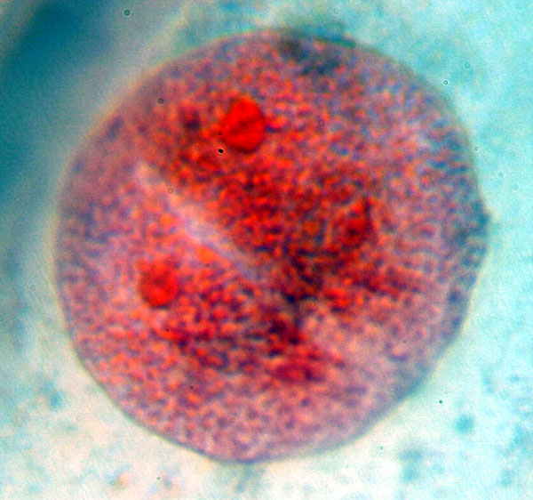
Why Acetocarmine is Used in Mitotic Chromosome Studies – Pediaa.Com
Jaime’s Human Biology Blog: THE MICROSCOPE, CELLS, AND ORGANELLES LAB
Perspective – Brie Childress, LISW-CP – Anderson, SC
Onion Cell Under Microscope 40x Labeled – Micropedia
Mitotic Cells Under The Microscope. Onion Root Tip Stock Photography | CartoonDealer.com #175412738
Onion cell | Teaching science, Cell, Microbiology
How To See Plant Cell Under Microscope : Biology Students observing plant and animal cells under …
The Wonderful Microworld: Onion cells Near the Skin Area
Cell-fie! Cheek Cell Lab | Gracyn’s Blog
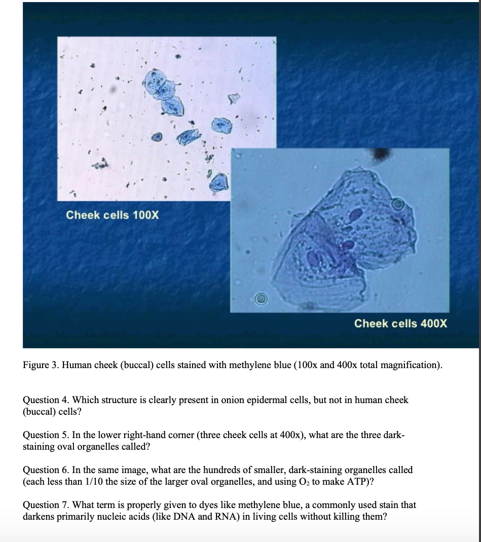
Solved Cheek cells 100X Cheek cells 400X Figure 3. Human | Chegg.com
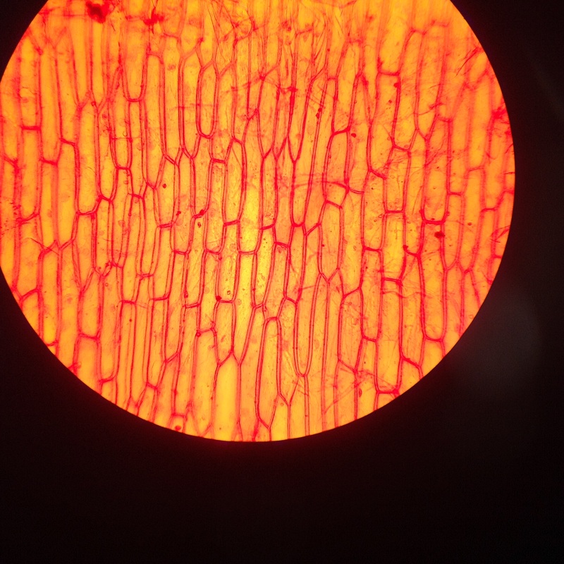
RED ONION; EPIDERMAL CELLS SHOWING CYTOPLASM AND NUCLEI; RED COLOR IS Stock Photo: 10349246 – Alamy
Onion Cells at the Microscope Stock Image – Image of background, division: 120810831
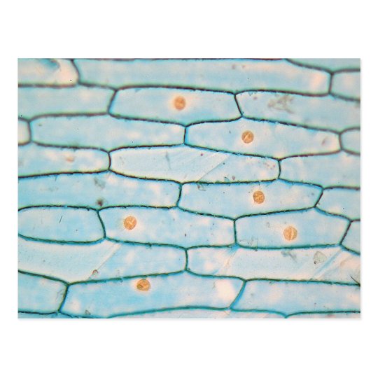
Onion under the microscope postcard | Zazzle.com
ONION SKIN CELLS (EPIDERMAL CELLS) SHOWS CELL STRUCTURE AND NUCLEUS STAINED IN IODINE / LIVE …
My Cheek Cells | 7th Grade Science
Microscopes TrueVision » Blog Archive 40X – 1600X TRINOCULARCOMPOUND LIGHT MICROSCOPEKOEHLER …
Calculated Images: Cheeky
Pin by Amy H on Scientific Drawing | Pinterest | Scientific drawing, Drawings and Biology
ONION SKIN CELLS EPIDERMAL CELLS SHOWS CELL STRUCTURE AND NUCLEUS STAINED IN IODINE LIVE 100X …
Onion cell(nucleus)
Through a Microscope stock photo. Image of cells, onion – 57855662
Onion Cells stock image. Image of cellular, microscope – 12996999
Prokaryotic & Eukaryotic Biological Cell Images & Photos
Calculated Images: Cheeky
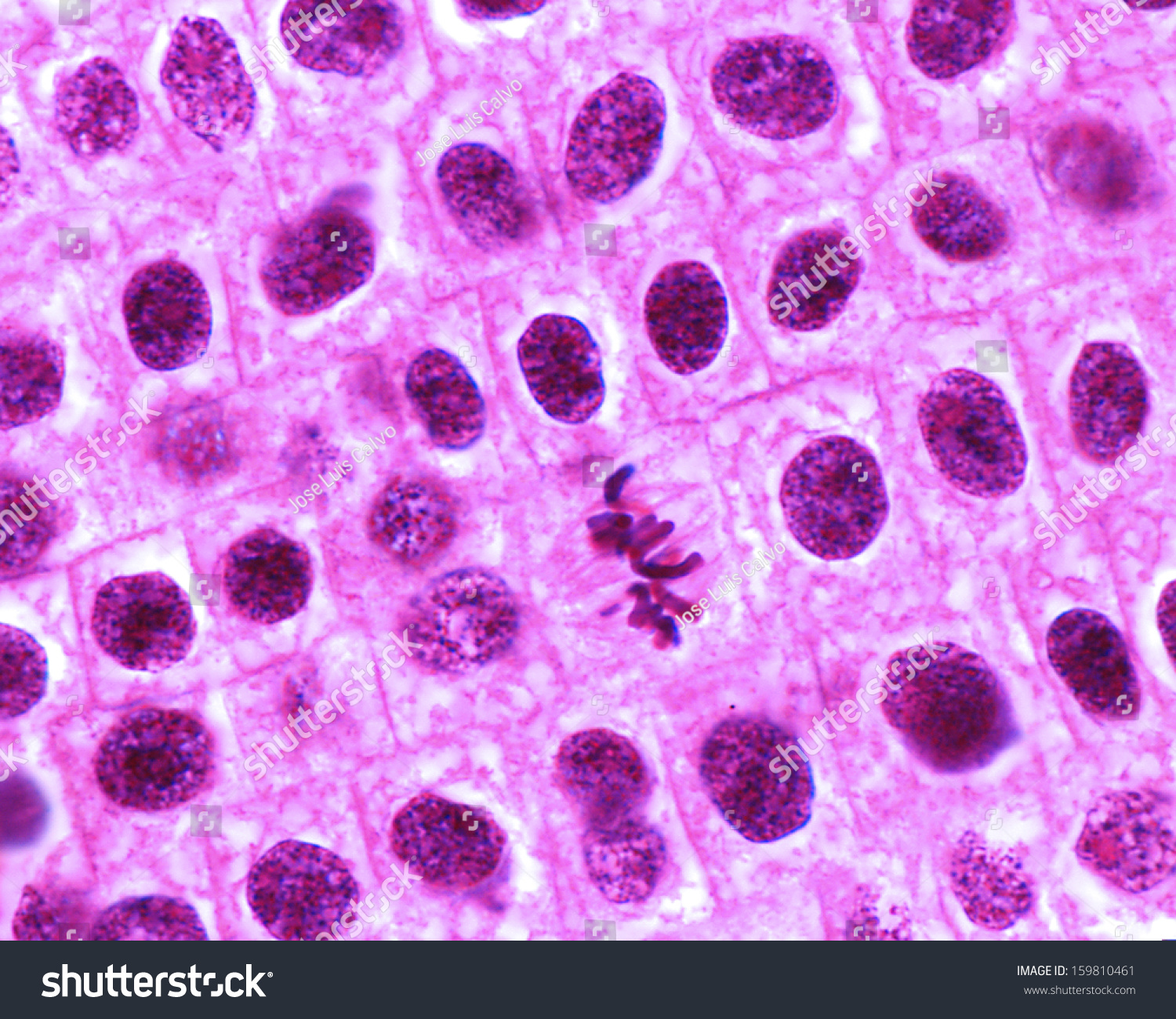
Mitosis In Onion Cells Of The Root Meristem. In The Center A Typical Metaphase Can Be Seen, With …
19 Parts Of A Compound Microscope Worksheet / worksheeto.com
We extend our gratitude for your readership of the article about
jack is seeing an onion cell under a microscope at
galleryz.online . We encourage you to leave your feedback, and there’s a treasure trove of related articles waiting for you below. We hope they will be of interest and provide valuable information for you.


