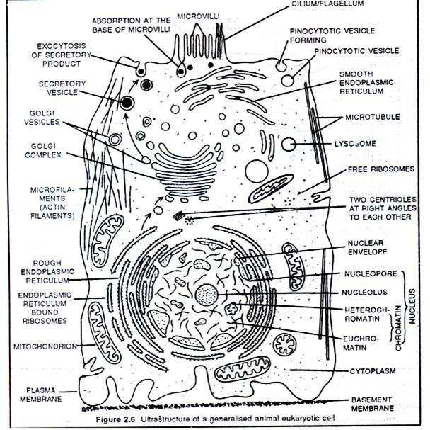
Solved Label the transmission electron micrograph of the | Chegg.com
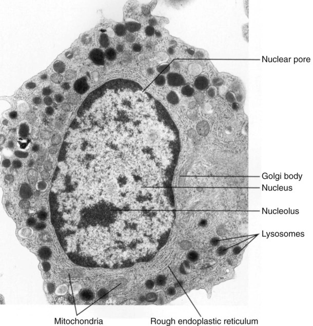
34 Label The Transmission Electron Micrograph Of The Nucleus – Labels 2021
ExcFIG 4 Legend
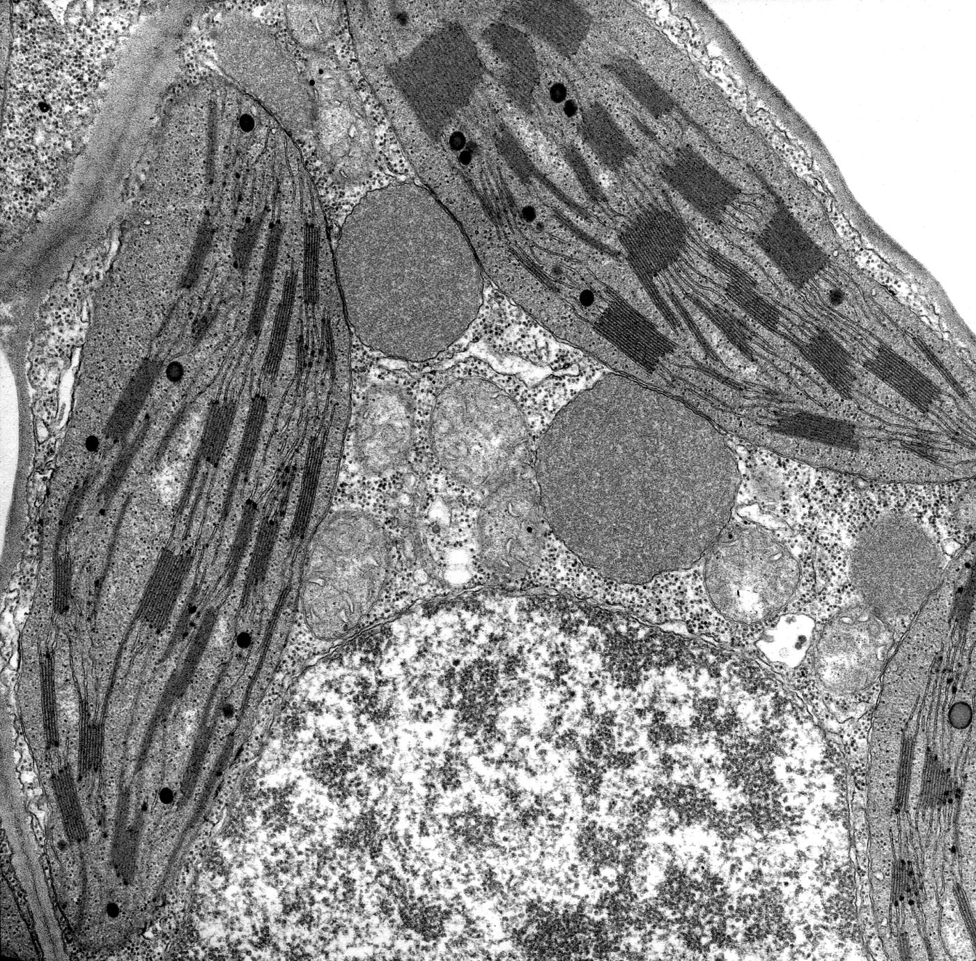
Transmission electron microscope image of a cell with ultrastructural …
33 Label The Transmission Electron Micrograph Of The Nucleus – Label …
33 Label The Transmission Electron Micrograph Of The Nucleus – Labels …
Transmission electron microscopy of a human eosinophil. This cell is …
Transmission electron microscopy of intracellular stages Rhytidocystis …
Transmission electron micrographs showing phloem cells of various types …
Transmission electron micrograph (TEM) of a section through an algal …
The morphology of mitochondria. (a) Thin-section electron micrograph of …
Transmission electron micrographs showing the morphology of untreated …
Transmission electron micrograph of a thin section through the …
Transmission electron microscopy showing the presence or near absence …
Transmission electron micrographs of representative glomerular …
Transmission electron micrograph of a Rickettsia sp. showing three …
Transmission electron microscopy (8.400x) of yeast cell wall …
Cell Membrane Micrograph High Resolution Stock Photography and Images …
lab3exercise
Transmission electron micrographs of mitochondria in equine oocytes. A …
Transmission electron micrograph of a cell of strain SVX8 T : ultrathin …
Transmission electron micrographs of blood cells in large Indian civet …
Transmission electron micrograph of human Sertoli cells in culture. A …
Transmission electron micrograph of a red blood cell Stock Photo …
Transmission electron microscopy of the cell envelope of mycobacteria …
34 Label The Transmission Electron Micrograph Of The Nucleus – Labels 2021
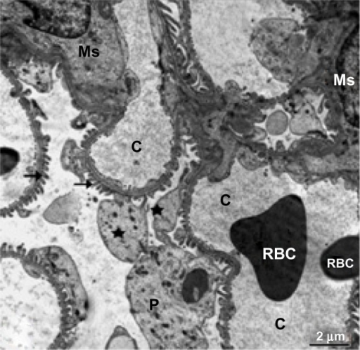
-Transmission electron micrograph of glornerular capillary loops. The …
Cellular Structure and Function | Oncohema Key
Transmission electron microscopy images of bacteria observed in the …
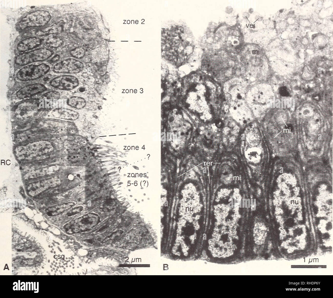
-Transmission electron micrograph of region I of the inner medulla (IM …
Transmission electron microscopy of the syncytiotrophoblast (ST) cell …
Transmission electron micrograph of transverse sections of Arabidopsis …
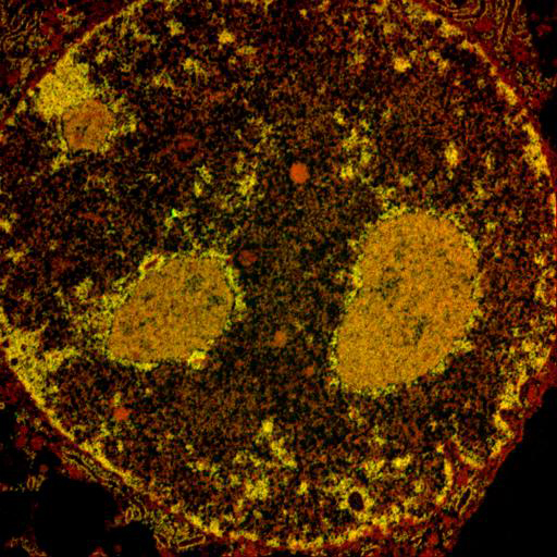
Nucleus Micrograph Gallery
Pin by NIA on Education | Plant cell, Electron microscope, Cell
Transmission electron micrographs of cells and scales of undescribed …
31 Label The Transmission Electron Microscope Image Of A Chloroplast …
Transmission electron microscopy images of immunogold-labeled secretory …
33 Label The Transmission Electron Micrograph Of The Nucleus – Labels …
33 Label The Transmission Electron Micrograph Of The Nucleus – Label …
24 best images about Transmission Electron Microscopy on Pinterest …
Transmission electron microscopy (TEM) images of the mesophyll cells of …
Transmission electron micrographs of BHV-1 entering MDBK cells. Panels …
Ultrasound-Mediated Therapy – Are We There Yet? | National Institute of …
Transmission electron micrographs of gram-negative prokaryotes within …
Label Electron Micrograph Plant Cells
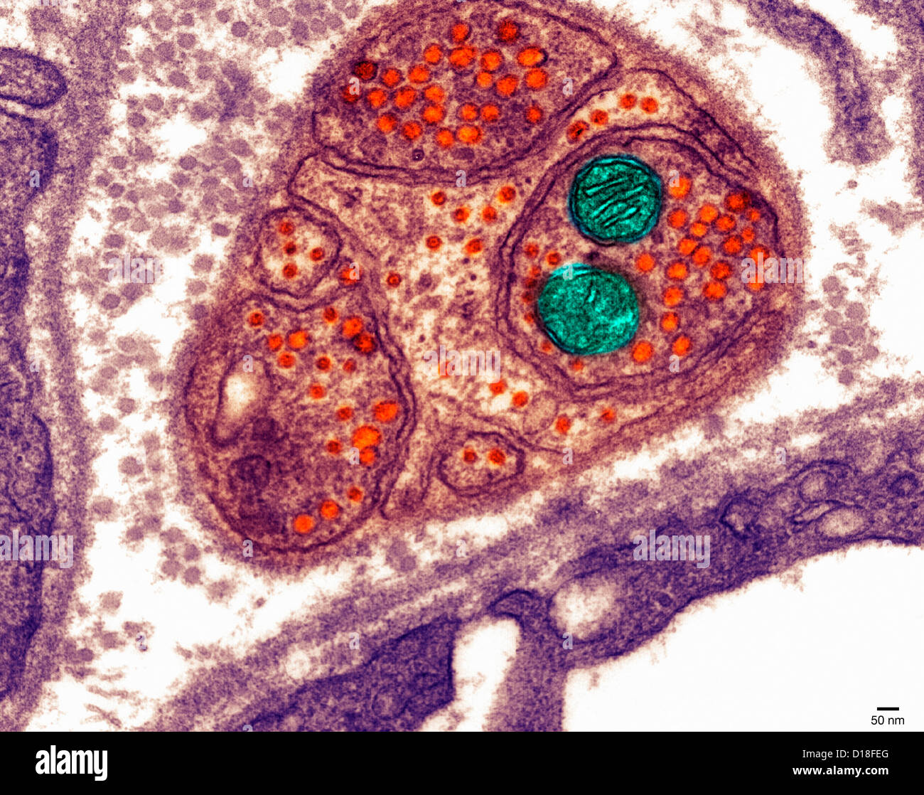
Nucleus, glyoxisomes, chloroplasts, and mitochondria – magnification …
33 Label The Transmission Electron Micrograph Of The Nucleus – Labels …
Transmission electron micrograph of a vesicle expelled from A …
Transmission Electron Micrographs (TEM) of mesophyll cells from tef …
Transmission electron micrograph of a thin section of P. acnes at 60 K …
Animal Cell Under Transmission Electron Microscope / Cell Organelles …
Transmission electron microscopy of N-cadherin CKO hearts. Electron …
Electron microscope, Scanning electron microscope, Electrons
Transmission electron micrograph of (A) conventional and (B …
31 Label The Transmission Electron Microscope Image Of A Chloroplast …
Transmission electron microscopy of A549 cells treated with LPZ or AZM …
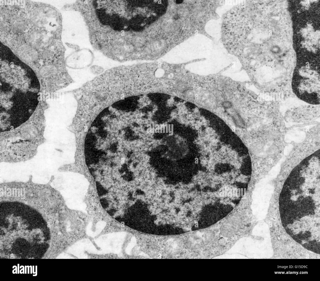
Electron micrograph demonstrating chromatin bodies (Ch) in the nucleus …
Transmission electron microscopy. Both cultures showed typical round or …
Transmission electron microscopy (TEM) of white blood cells in a …
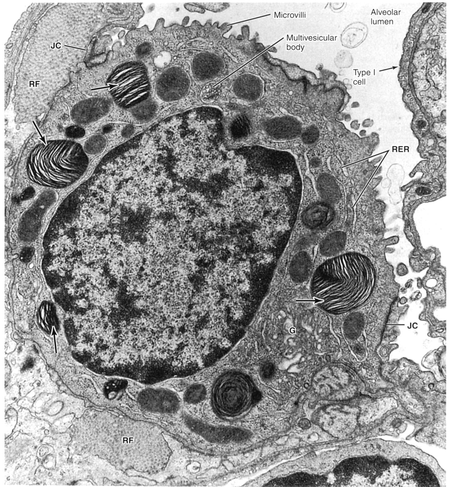
Transmission electron micrograph of normal canine lung. The flattened …
Transmission electron microscopy of a specimen taken from a patient …
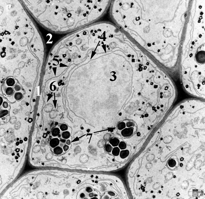
Transmission electron micrograph of kidney glomerular t | Open-i
Electron Microscope: Definition, Types, Parts, Application, Advantages …
Animal Cell Diagram Electron Microscope : Labeled Plant Cell Under …
-Transmission electron micrograph of thin limbs of Henle’s loop. (a …
Transmission electron microscopy of ischemic (a) and… | Download …
Search in gallery
Transmission electron micrograph of two endosymbionts in an I …
Transmission electron microscopy images showing details of the muscle …
Transmission electron micrograph of transverse sections of Arabidopsis …
Transmission electron microscopy of peripheral blood DC subsets. TEM …
Lab Section Number – Applied Botany – Florence Grovida Gardening
Transmission electron micrographs of bacterial cells infected by …
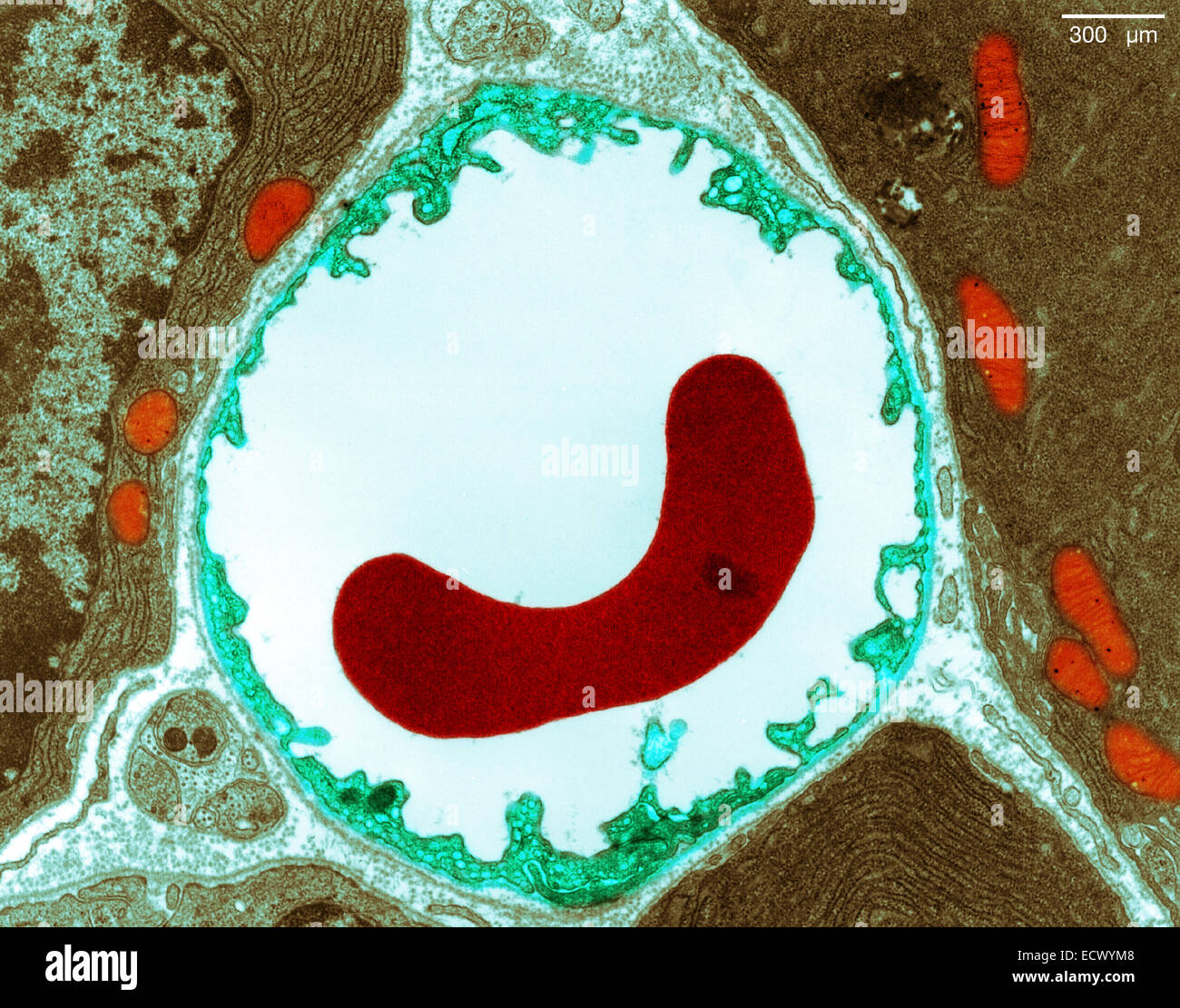
Transmission electron micrograph (TEM) of a section of blood vessel …
Human neutrophil cell tem hi-res stock photography and images – Alamy
Transmission Electron Microscopy Gallery – Center for Microscopy and …
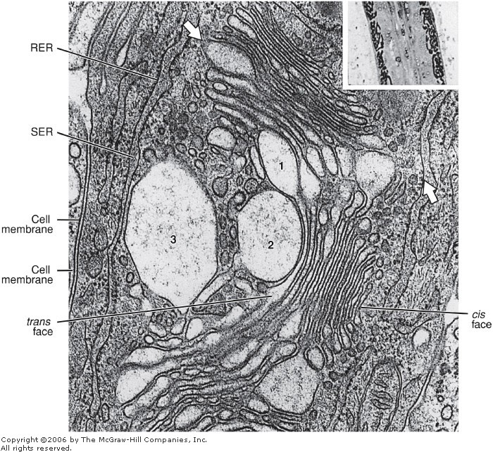
De Histology: Golgi Complex (Golgi Apparatus)
Electron microscopy – Auditory Science lab
Representative electron micrograph showing ultrastructure of cells form …
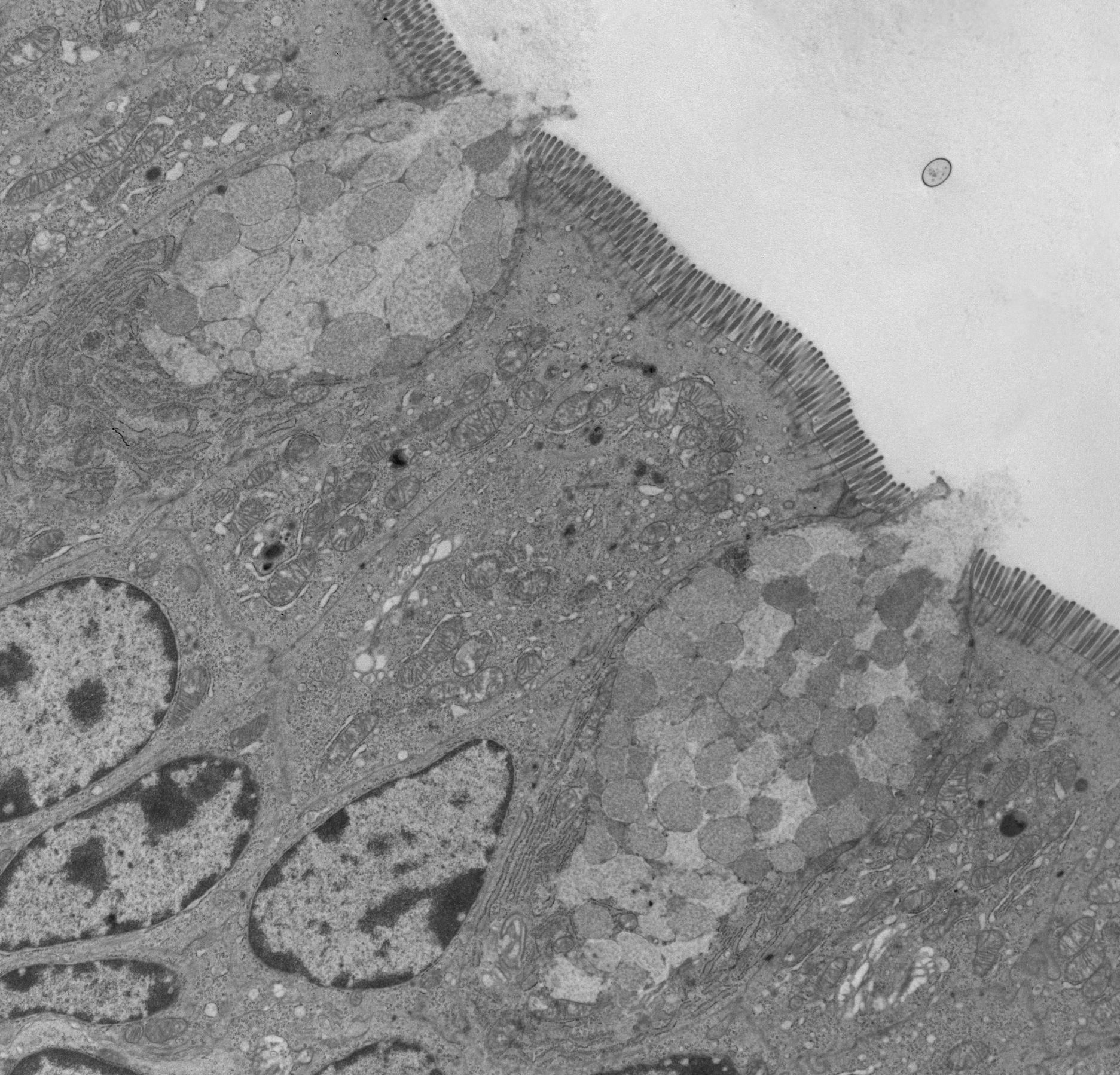
Electron Micrograph Showing Parts of Two Epithelial Cells of a …
Transmission electron microscopy images of secretory vesicles in growth …
Transmission electron micrographs of liver sections from Wt and Atgl−/− …
Transmission electron micrograph of intact bacteria (arrows) directly …
Transmission electron microscopy (TEM) of tissue formed by hP2bP0 …
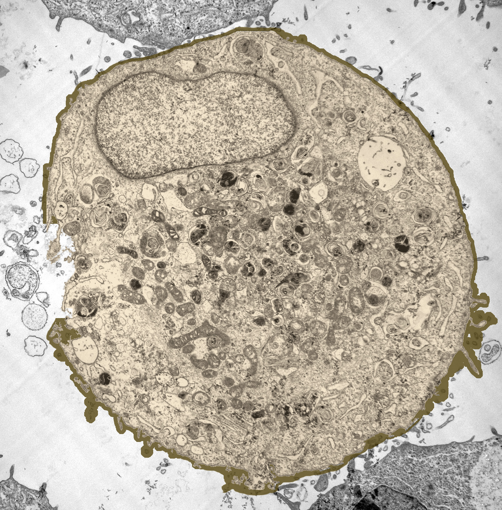
Electron Micrograph
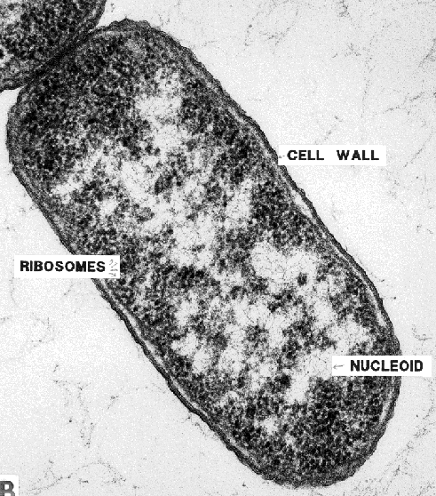
More on Bacterial Morphology
Transmission Electron Microscopy (TEM) images: intact yeast cells of H …
33 Label The Transmission Electron Micrograph Of The Nucleus – Label …
Transmission electron micrograph of a liver cell (72/22) cloned from a …
2. Transmission electron micrographs of the nuclear envelope …
Transmission electron microscopy of the endosperm from the double …
Research | Molecular Cell Biology
Transmission electron micrograph of B16 melanoma cells, with and …
Transmission Electron micrograph of a thin transversal cut through a …
Transmission electron microscopy showing a mesothelioma cell cultured …
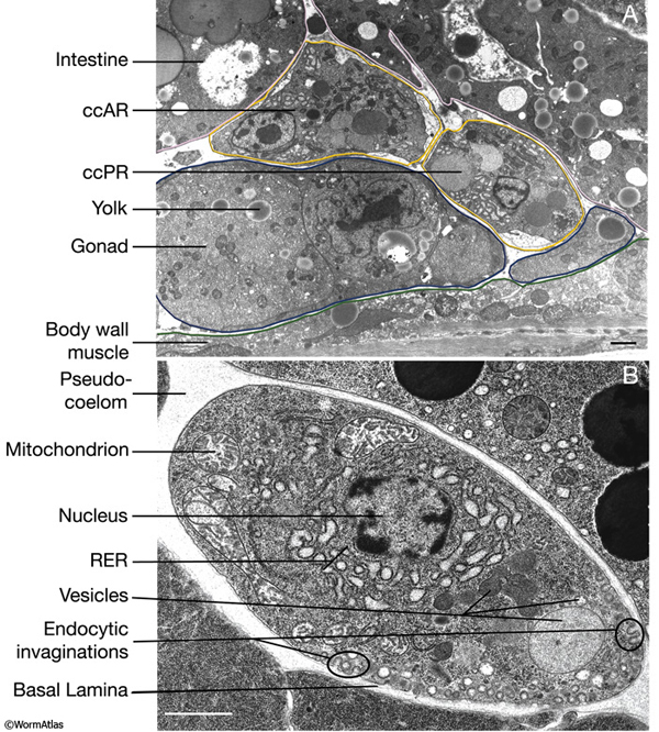
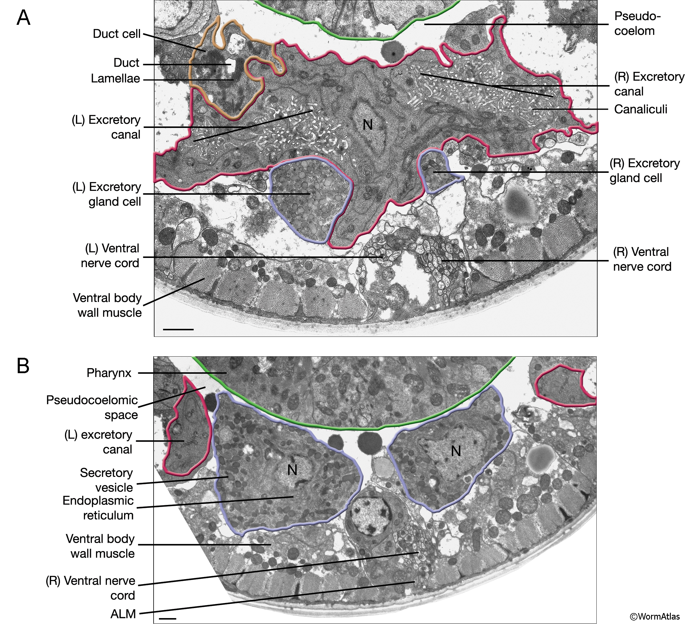
Ben Mulcahy 2023 C. elegans Meeting Workshop
We extend our gratitude for your readership of the article about label the transmission electron micrograph of the cell at galleryz.online. We encourage you to leave your feedback, and there’s a treasure trove of related articles waiting for you below. We hope they will be of interest and provide valuable information for you.