List showcases captivating images of radiography of the uterus and oviducts with contrast medium galleryz.online
radiography of the uterus and oviducts with contrast medium
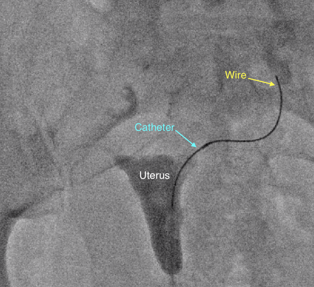
Fallopian Tube Blockage / Recanalization Doctor – Los Angeles, California
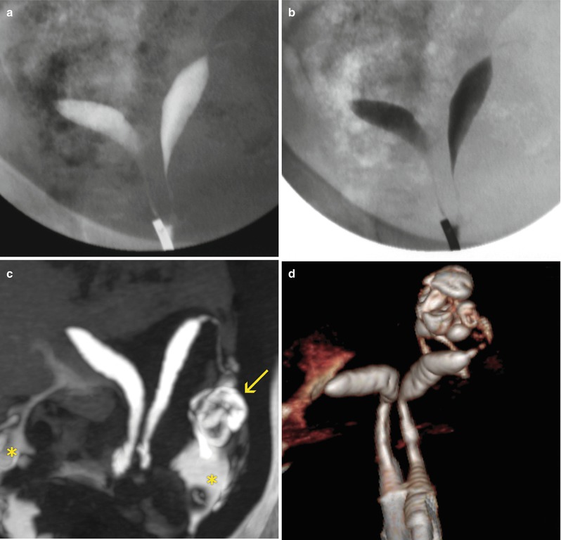
Normal or Abnormal? Demystifying Uterine and Cervical Contrast …
Comparison of uterine lesions. (A) Pelvic magnetic resonance imaging …
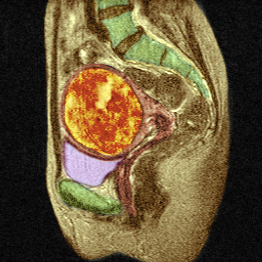
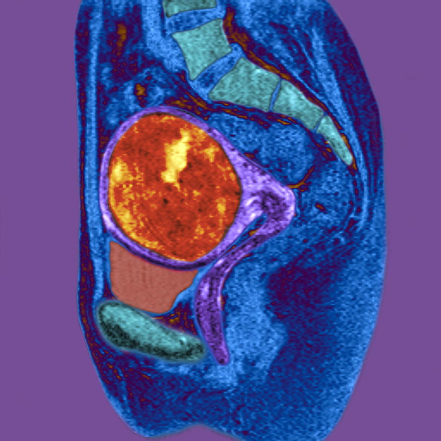
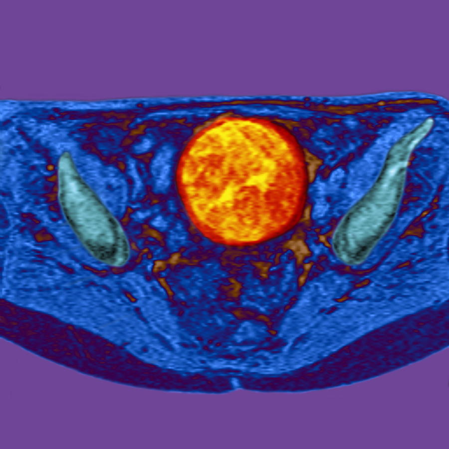
Uterine Fibroid, Mri Scan Photograph by Du Cane Medical Imaging Ltd
Uterine Fibroid, Mri Scan Photograph by Du Cane Medical Imaging Ltd
Uterine Wall Pathology | Radiology Key
Management of Uterine Fibroids: A Focus on Uterine-sparing …
MRI appearances of benign uterine disease – Clinical Radiology
Uterine Fibroid, Mri Scan Photograph by Du Cane Medical Imaging Ltd
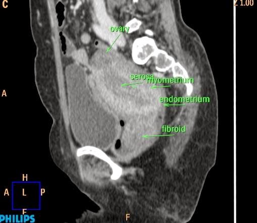
(A) Computed tomography shows 14.2 cm huge uterine myoma with …
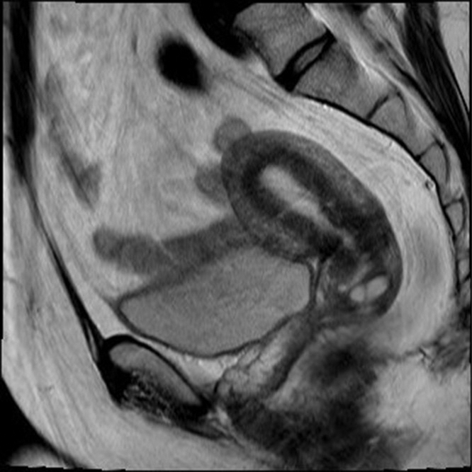
Pelvic MRI showing a normal uterus (A), and about a 7.5×6.2×5.8 cm …
Normal or Abnormal? Demystifying Uterine and Cervical Contrast …
Adnexal Torsion: Review of Radiologic Appearances | RadioGraphics
MR Imaging of the Uterine Cervix: Imaging-Pathologic Correlation …
Dynamic MR Imaging of the Pelvic Floor: a Pictorial Review | RadioGraphics
Pseudo aneurysm of the uterine artery with arteriovenous fistula after …
Adenomyosis of the uterus | Image | Radiopaedia.org
Adenomyosis of uterus | Image | Radiopaedia.org
Imaging studies. (A) Uterus and hemoperitoneum transvaginal ultrasound …
UTERINE ULTRASOUND IMAGING
Imaging tests of the uterine cervix. (A and B) The ultrasound scanning …
Figure 1 from Magnetic Resonance Imaging and Ultrasound Depiction of …
Spectrum of CT Findings in Acute Pyogenic Pelvic Inflammatory Disease …
Post Partum Uterus / CTisus.com | Pediatrics, Uterus, Case study
Uterus didelphys | Image | Radiopaedia.org
Additional pathologies found during uterine fibroid MRI screening. a …
UTERINE ULTRASOUND IMAGING
UTERINE ULTRASOUND IMAGING
Normal or Abnormal? Demystifying Uterine and Cervical Contrast …
Top: Partial septate uterus. The fundal line measure is > 5 mm as …
Three-dimensional ultrasound imaging of the normal uterus in coronal …
An ultrasound picture of an adenomyotic uterus shows the characteristic …
Benign gynecologic lesions: Vulva, Vagina, Cervix, Uterus, Oviduct …
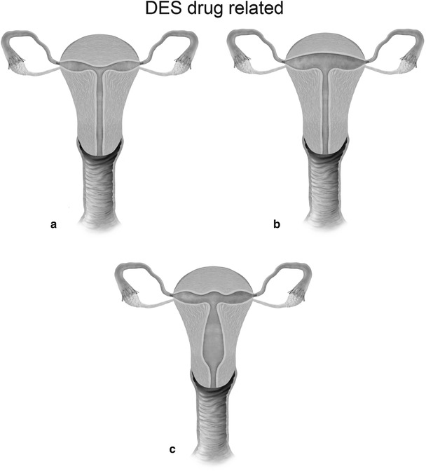
Deformed uterus, X-ray – Stock Image M850/0516 – Science Photo Library
Magnetic Resonance Imaging in Gynecology | GLOWM
Top 11 Doctor Insights on thickened endometrium ultrasound – HealthTap …
Didelphys uterus (A) schematic representation; (B) spot radiograph of …
Translabial US and Dynamic MR Imaging of the Pelvic Floor: Normal …
(PDF) What Is the Role of Imaging at Primary Diagnostic Work-Up in …
Pin on Ultrasound uterine Anatomy
Phlebography; Venography
Sonographic findings of early pregnancy in the rudimentary horn of a …
Journal of Postgraduate Gynecology & Obstetrics: October 2014
UTERINE ULTRASOUND IMAGING
Robert’s uterus: modern imaging techniques and ultrasound‐guided …
Bicornuate uterus in teenager. Transvaginal US. A, Transverse plane …
-CT transversal imaging of abscess formation in front of uterus. A …
Uterine fibroid, MRI scan – Stock Image – M850/0557 – Science Photo Library
Congenital Uterine Anomalies | Radiology Key
Multiplanar imaging of a normal uterus at volume ultrasound: the volume …
Large lower uterine segment mass.T2 sagittal pelvic MRI with contrast …
Normal or Abnormal? Demystifying Uterine and Cervical Contrast …
Transabdominal ultrasound of the uterus. Note the urinary bladder …
Pin on Radiología
MR Imaging of the Uterine Cervix: Imaging-Pathologic Correlation …
Uterus didelphys | Image | Radiopaedia.org
Diffusely Enlarged Uterus: Evaluation with MR Imaging | RadioGraphics
Effect of Uterine Artery Embolization on Uterine and Leiomyoma …
Hysterosalpingogram demonstrates a communicating septate uterus, cervix …
UTERINE ULTRASOUND IMAGING
(A) Transvaginal ultrasound Doppler image showed a giant mass …
MR imaging of IUD. A-C. Images of uterus of 42-year-old woman with …
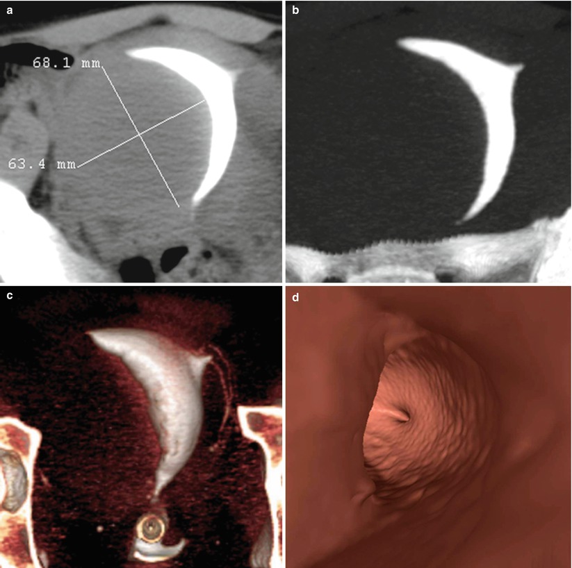
Plain CT showing barium contrast within the uterus and pouch of …
Ultrasonographic section of the uterine body/cervical leiomyoma that is …
US of the Pediatric Female Pelvis | Radiology
Effect of magnetic resonance imaging characteristics on uterine fibroi …
WK 3 L 2 Intramural fibroid | Diagnostic medical sonography, Ultrasound …
OB/Gyn Images | Ultrasound, Transvaginal ultrasound, Medical illustration
MR Imaging of the Uterine Cervix: Imaging-Pathologic Correlation …
(Top) Laparoscopic identification of a bicornuate uterus with a dilated …
Hypercellular uterine leiomyoma: MRI findings | Eurorad
Uterine Fibroids, Mri Scan Photograph by Zephyr
(a) TA Sag Uterus. Transabdominal sagittal view of the uterus shows …
Endometrial/junctional zone thickening | Radiology Key
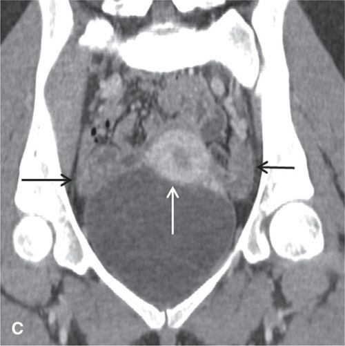
Contrast-enhanced computed tomography image showing displaced uterus to …
JCI – Tuning the oviduct to the anandamide tone
Diagnostic Imaging | Obgyn Key
Cross-Sectional Imaging of the Uterus | Radiology Key
Effect of magnetic resonance imaging characteristics on uterine fibroi …
Ultrasound Evaluation of the Uterus | Obgyn Key
Normal or Abnormal? Demystifying Uterine and Cervical Contrast …
Complete obliteration of the uterine cavity. Extrauterine intravasation …
Magnetic resonance imaging of the pelvis showing normal ovaries (thick …
Uterine growth, ultrasound scans – Stock Image – M850/0488 – Science …
Coronal reformat image of a contrast-enhanced Abdomen and Pelvis CT …
MR imaging of bladder endometriosis and its relationship with the …
Incarceration of the gravid uterus: diagnosis and preoperative …
(PDF) Imaging features of postpartum uterine rupture: A case report
MR Imaging of the Uterine Cervix: Imaging-Pathologic Correlation …
MR Imaging of the Uterine Cervix: Imaging-Pathologic Correlation …
Preoperative computed tomography scans of a huge fibromatous uterus …
(PDF) Contrast-Enhanced Dynamic MR Imaging of Uterine Fibroids as a …
Magnetic resonance imaging of pelvis demonstrates uterus didelphys with …
MR Imaging-based Assessment of the Female Pelvic Floor | RadioGraphics
Uterine Arteriovenous Malformations – O’Brien – 2006 – Journal of …
