Collection showcases captivating images of the area of the sarcomere where the actin and myosin myofilaments overlap is called the gathered and meticulously curated by the website galleryz.online. Furthermore, you can find more related images in the details below.
the area of the sarcomere where the actin and myosin myofilaments overlap is called the
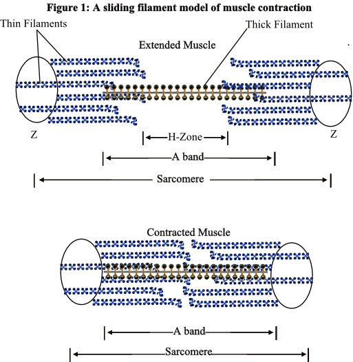
 Sarcomere muscular biology scheme vector illustration Easy Science …
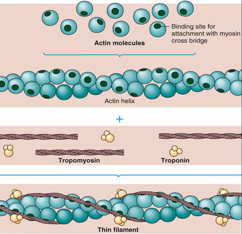
 Actin and Myosin | Biology Dictionary
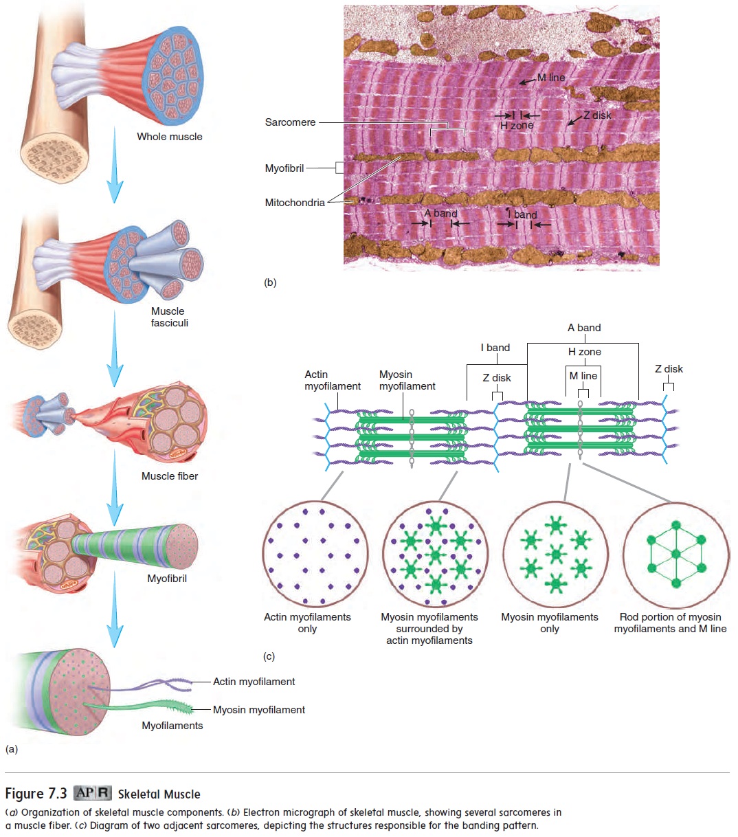
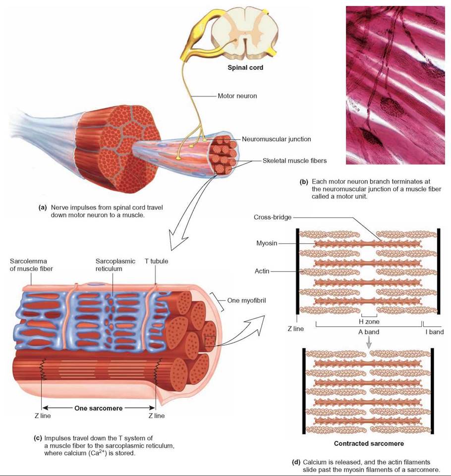
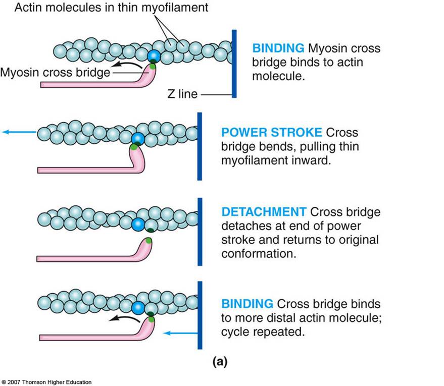
 10.2 Skeletal Muscle – Anatomy & Physiology
 10.2 Skeletal Muscle – Anatomy and Physiology
 Weight Training 101: How Muscles Work — EvanTraining
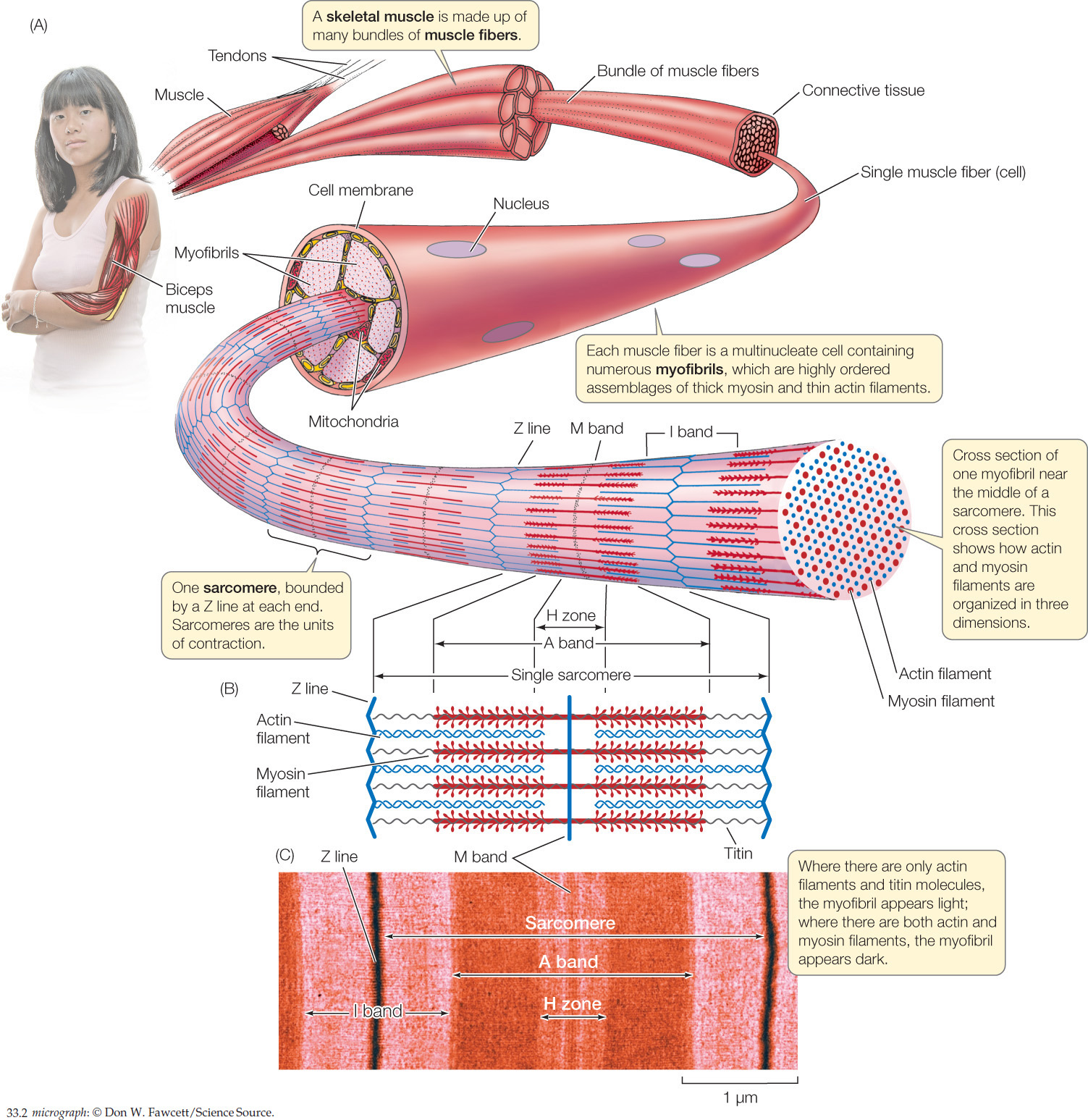 hillis2e_ch33
 Chapter 9: Muscles and Muscles Tissue | HNFE 3804: Ex Phys | Pinterest …
 Human Physiology – Muscle | Physiology, Medical school essentials …
 Muscle – Medical Biochemistry
 Sarcoplasmic Hypertrophy | Skeletal muscle, Muscle hypertrophy, Science …
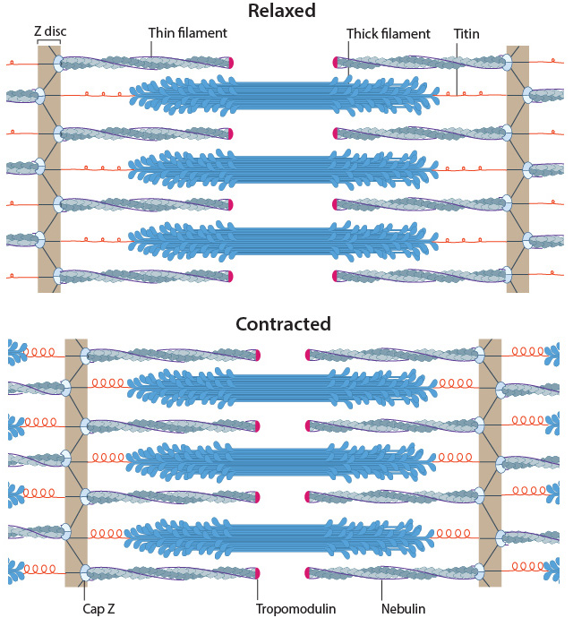
 Structure and position of titin in a sarcomere. | Human anatomy and …
 Anatomy of a skeletal muscle and a sarcomere. (A) From SEER training on …
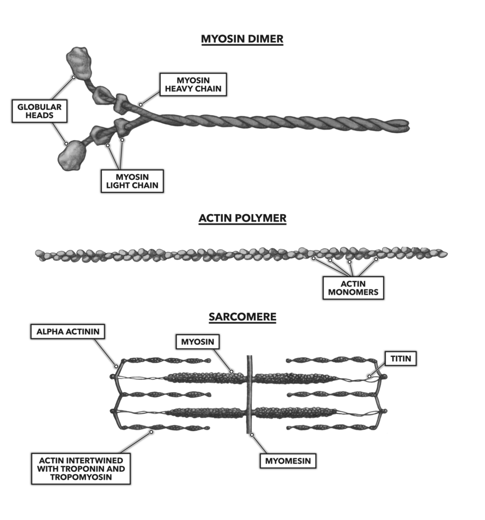
 The location and arrangement of the most abundant proteins in the …
 【myosin】什么意思_英语myosin的翻译_音标_读音_用法_例句_在线翻译_有道词典
 Titin location and arrangement in the cardiac sarcomere. | Download …
 (a) Electron micrograph of a vertebrate striated muscle sarcomere (here …
 Cardiac myosin binding protein-C as a central target of cardiac …
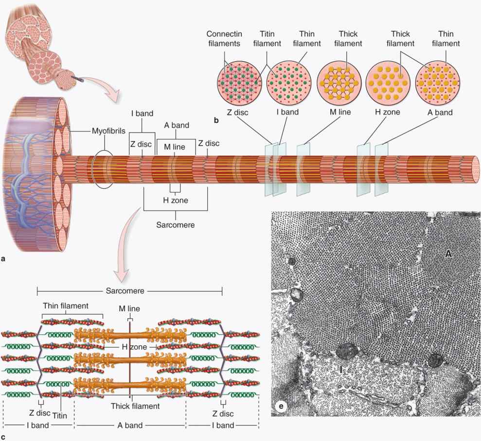
 Muscular Levels of Organization | Anatomy and Physiology I
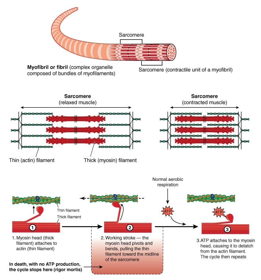 What Is Rigor Mortis? How Long Does Rigor Mortis Last?
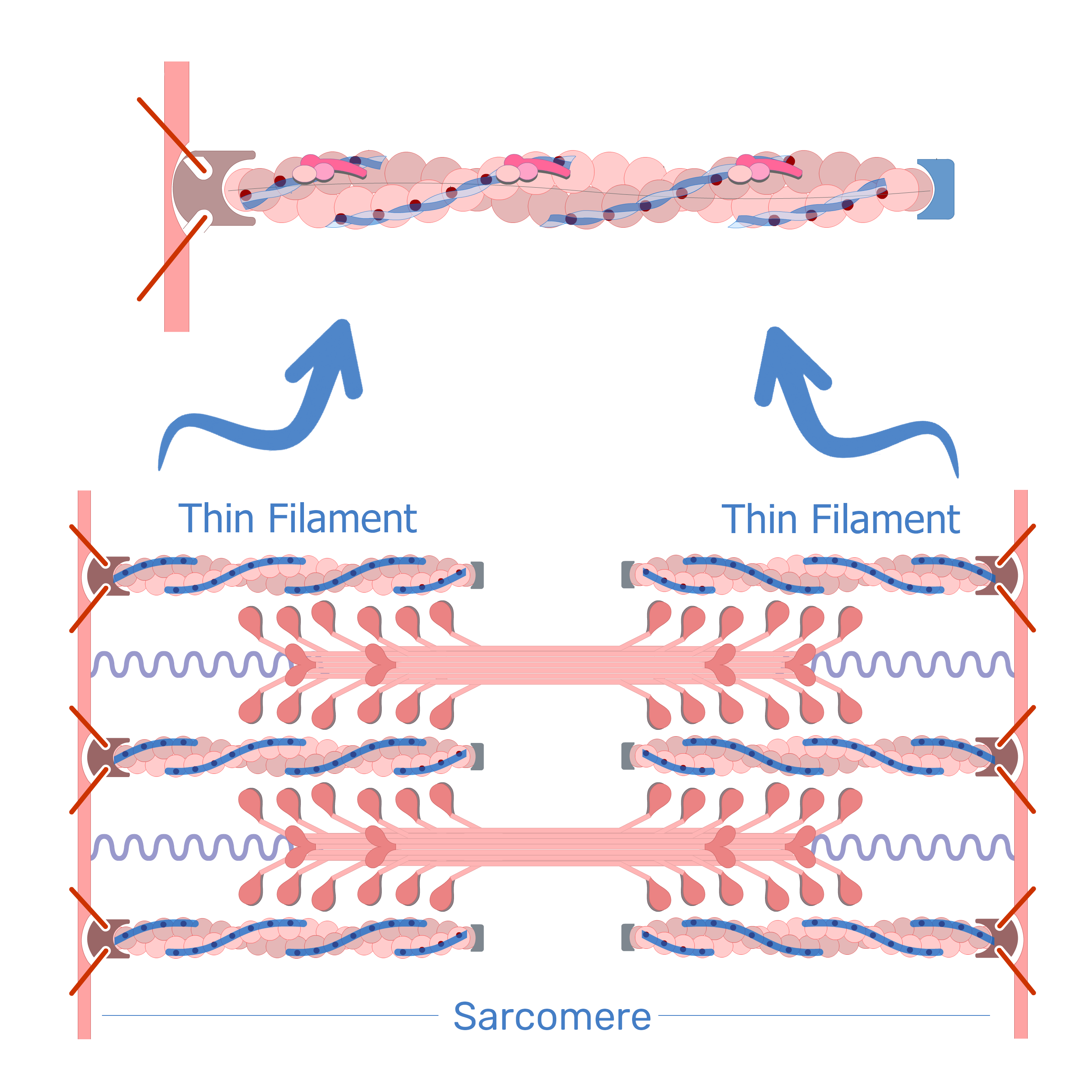 Thin filaments: definition, composition and function | GetBodySmart
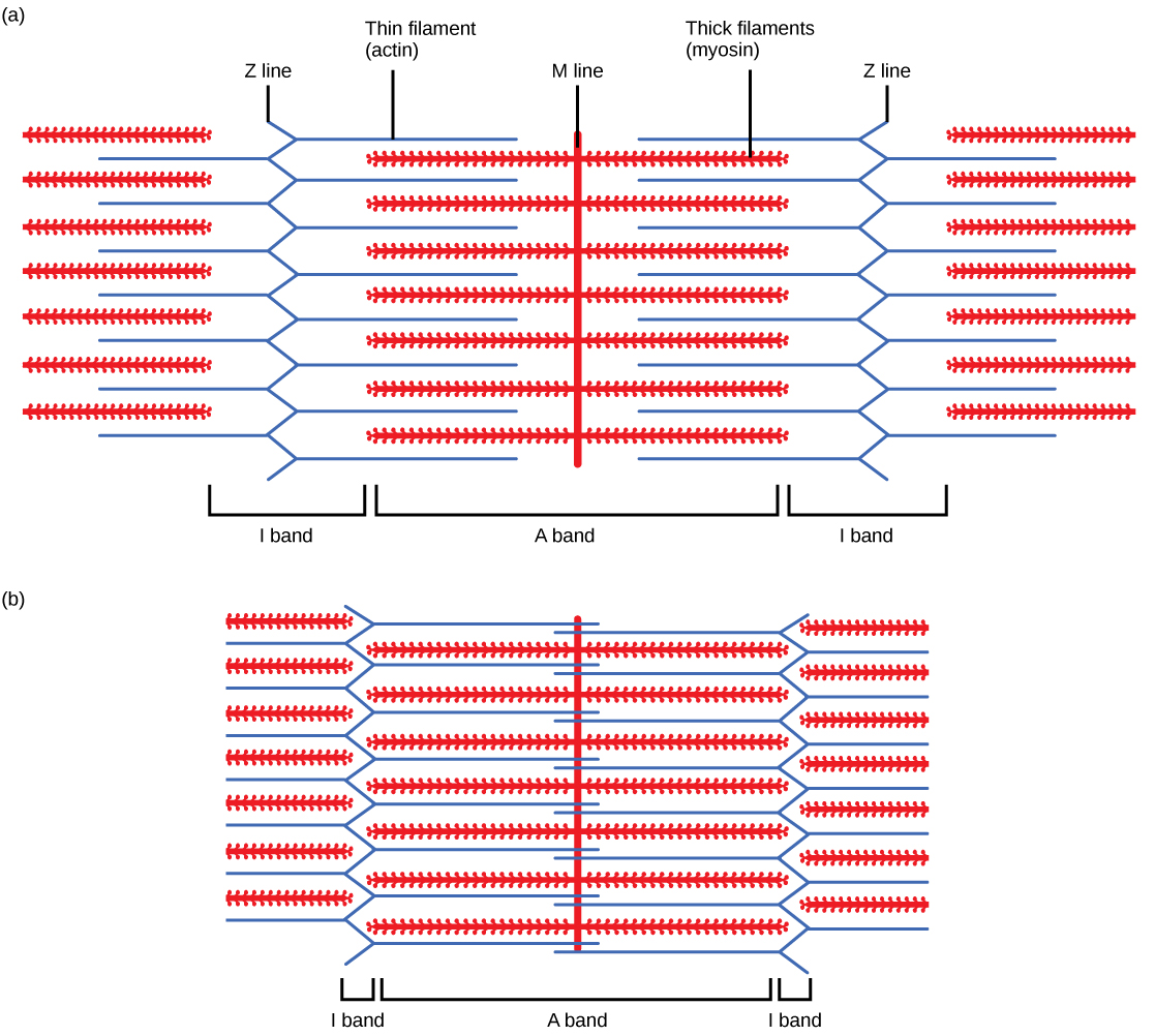
 Identify The Structures Labeled A B And C In The Diagram Of A Sarcomere …
 6.4: Muscle Contraction – Medicine LibreTexts
 Structure of the Sarcomere the Contractile Unit of Skeletal Muscles …
 Actin and Myosin | Biology Dictionary
 Sketch of a sarcomere, thick filament and a thin filament structure …
 (A) Schematic representation of a cardiac sarcomere (lacking nebulin …
 Scheme of sarcomere structure. Packing of main muscle proteins in thin …
 A, schematic diagram of the sarcomere with thick (titin | Open-i
 How Muscles Work (Part 2 of 2) – ShapeLog
 (A) Schematic representation of the sarcomere. The actin thin filament …
 Major components of the cardiac sarcomere: Actin (green), myosin (red …
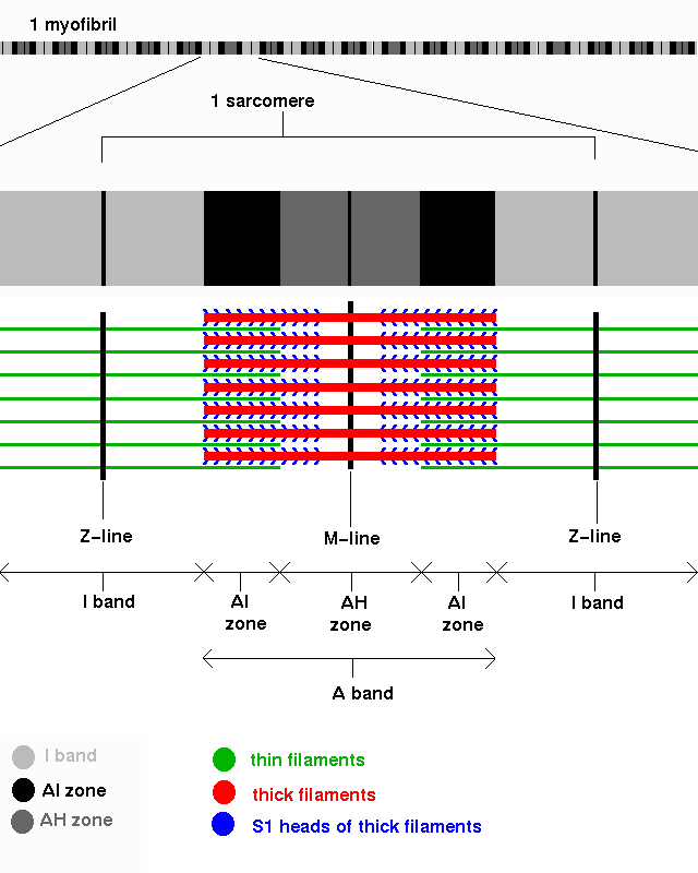
 4: Sarcomere organization in a myofibril. Adapted from Mesher AL …
 Posttranslational modifications of cardiac troponin T: an overview …
 Multiple Choice Questions on Muscle Contraction
 Schematic representation of sarcomere. (A). Sarcomeres are composed of …
 -Sarcomere consisting on a bundle of thin and thick myofilaments made …
 Sarcomere – Muscle Contraction
 Print Chapter 10 flashcards | Easy Notecards
 -Sarcomere consisting on a bundle of thin and thick myofilaments made …
 Simulation of stretching a single sarcomere beyond actin-myosin …
 Associate Degree Nursing Physiology Review
 Chapter 9-Muscles and Muscle Tissue Diagram | Quizlet
 What Is The Role Of Actin And Myosin – sharedoc
 Cardiac skeletal muscle sarcomere. Proteins localized in the cardiac …
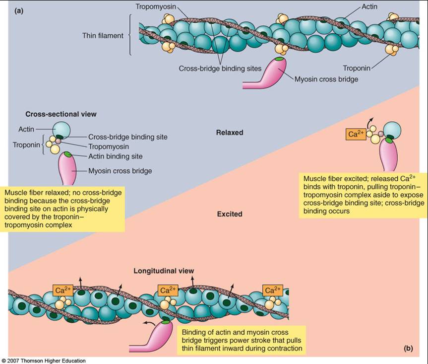
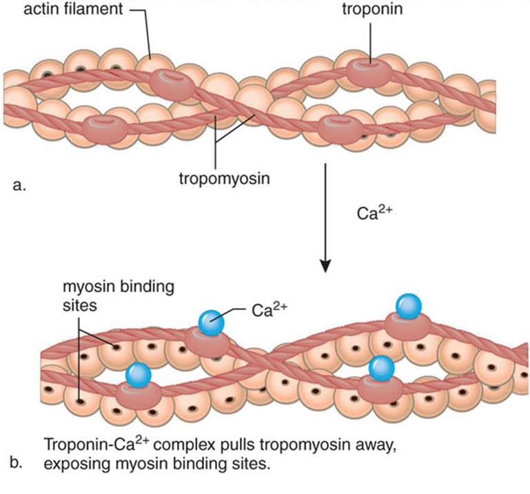
 (A) The active site on actin is exposed as calcium binds to troponin …
 Steps Of Muscle Contraction – Plasma Membrane
 Actin Cytoskeleton | Celebrate Cytochemistry | Gwen V. Childs, Ph.D.
 Actin Myosin Interaction
 Sarcomere-specific chaperones and ubiquitin ligases are necessary for …
 (PDF) Muscle contraction: Sliding filament history, sarcomere dynamics …
 Sarcomere mechanics in striated muscles: from molecules to sarcomeres …
 What is the difference between myosin and actin …
 What is Actin and Myosin? Easy Explanation of Movement
 PPS ’96: Muscle Fibres Part 2
 Structure of skeletal muscle myofibrils and role of the Z-disk …
 The relationship between the shortening of the sarcomere and the …
 Myosin VI Rewrites the Rules for Myosin Motors: Cell
 Schematic diagram of the sarcomere and costamere protein complexes of …
 Print The Muscular System flashcards | Easy Notecards
 Contractile Fiber
 Skeletal Muscle Structure
 Sarcomère : définition et explications
 Z-Line – AnatomyZone
 Nebulin is a large sarcomeric protein that is coextensive with the …
 IJMS | Free Full-Text | Myosin Assembly, Maintenance and Degradation in …
 Nature’s autonomous oscillators
 Identify The Structures Labeled A B And C In The Diagram Of A Sarcomere …
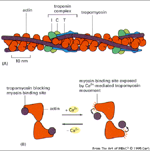
 10 Troponin regulation of actin-myosin interaction. Troponin I binds to …
 (a) Electron micrograph showing a whole sarcomere from fish muscle in …
 Scheme of sarcomere structure. Packing of main muscle proteins in thin …
 Myosin-V ATP cycle. (a) Both myosins bind ADP and are attached to actin …
 (PDF) Chapter 7 Molecular Structure of the Sarcomere
 Position of MyBPC in the stretched sarcomere and the structure of …
 Content Background: How Does the Alteration of Genetic Function by …
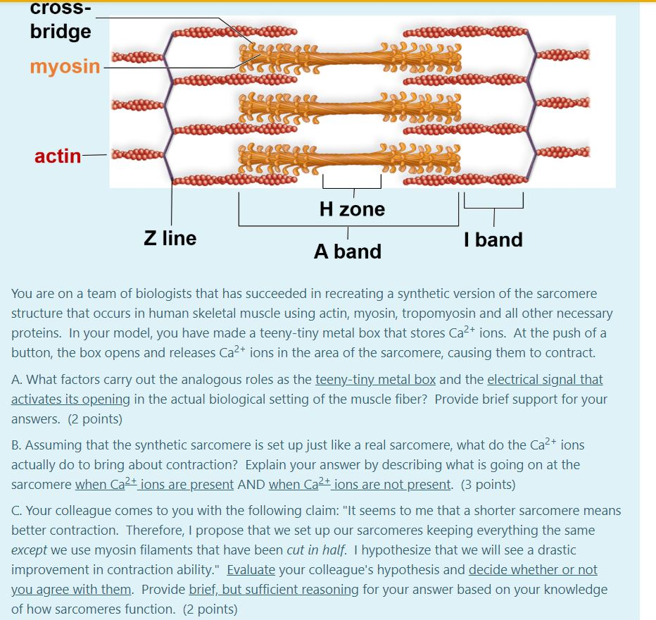 Solved blood cross- bridge myosin actin- H zone Z line I | Chegg.com
 Cells | Free Full-Text | Conventional and Non-Conventional Roles of Non …
 The kinesin-myosin walks. (a) Myosin motor mechanism. (i) Motor head …
 Cell types. Skeletal muscle cell. Atlas of Plant and Animal Histology.
 Regulation of muscle force in the absence of actin-myosin-based cross …
 Thin filament organization during sarcomere contraction in neonatal …
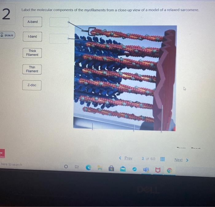 Solved Label the molecular components of the myofilaments | Chegg.com
 Muscle Cell (Myocyte): Definition, Function & Structure | Biology
 Output Coordination – The Body’s Control Mechanisms and Immunity …
 CrossFit | Muscle Basics, Part 1: Cells, Proteins, and Sarcomeres
 Chapter 9: Muscles and Muscles Tissue | Human anatomy and physiology …
 Muscle Fiber. 1. Myofibrils 2. Mitochondrium 3. Postsynaptic membrane 4 …
 Muscle Tissue | Basicmedical Key
 Associate Degree Nursing Physiology Review
 Solved: A typical relaxed sarcomere is about 2.3 μm in length a …
 Titin-based mechanosensing and signaling: role in diaphragm atrophy …
 Associate Degree Nursing Physiology Review
 Muscle Fibers. Fast Twitch Muscles. Building Muscles Fiber
 What is Actin and Myosin? Easy Explanation of Movement
 Myosin diagrams. (A) The classic myosin diagram pictures two equivalent …
 (A) The arrangement of myosin filaments in a straight myofibril. The …
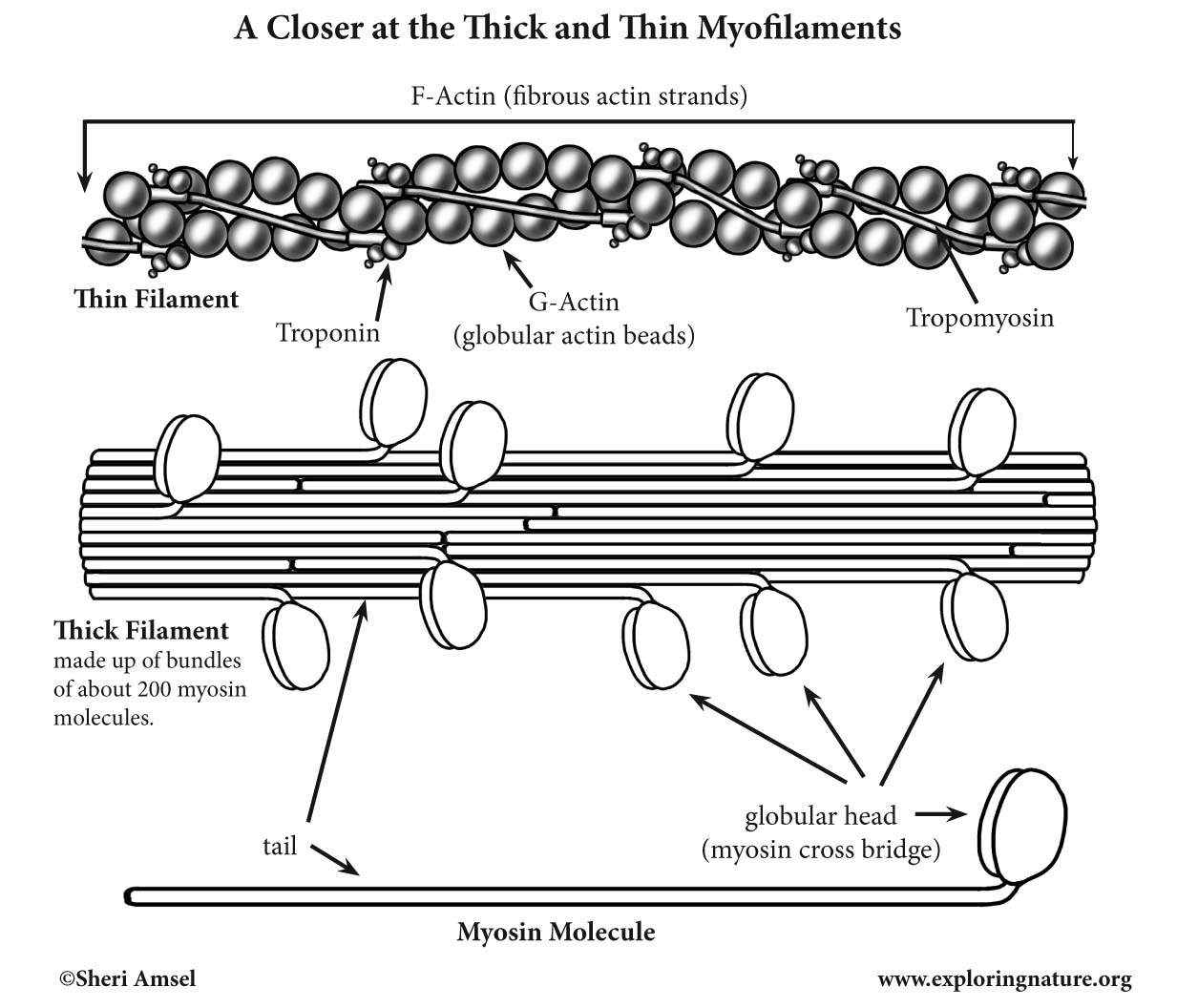 Muscle Myofilaments (Thick and Thin) (Advanced)
 A straightforward guide to the sarcomeric basis of cardiomyopathies | Heart
 Sarcomere thin filament disease candidates. Using the online database …
 (PDF) Sarcomere Structure: The Importance of Desmin Protein in Muscle …
 | Composite driven active rod experiments. (A) The rods are composed of …