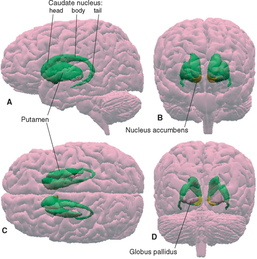
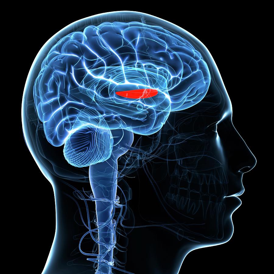
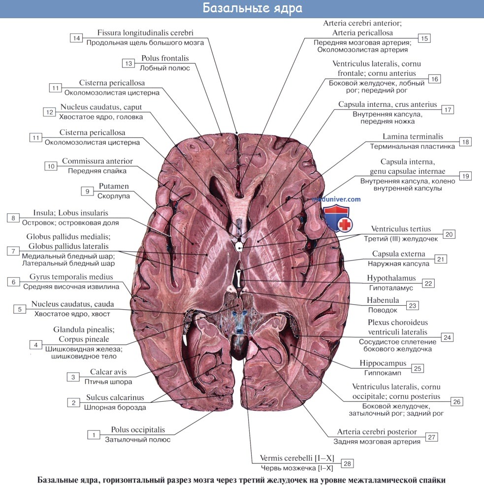
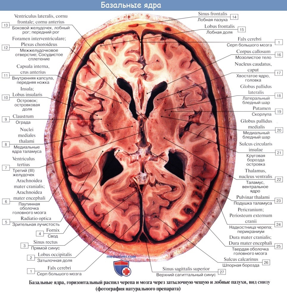
List showcases captivating images of the caudate putamen and globus pallidus compose the galleryz.online
the caudate putamen and globus pallidus compose the
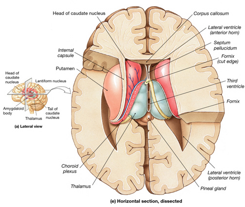
Depicts putamen, globus pallidus, thalamus, caudate nucleus, internal …
Putamen And Globus Pallidus Form | Globus
Globus Pallidus Putamen | Globus
Caudate Putamen Globus Pallidus | Globus
The Potential Role of the Striatum in Antisocial Behavior and …
Illustration of segmentation and the corresponding 3D structures in …
Pin on Anatomy
Coronal Section Through Cerebral Hemisphere | ClipArt ETC
3. The subcortical structures of the brain. Globus pallidus is called …
Insular Cortex Article
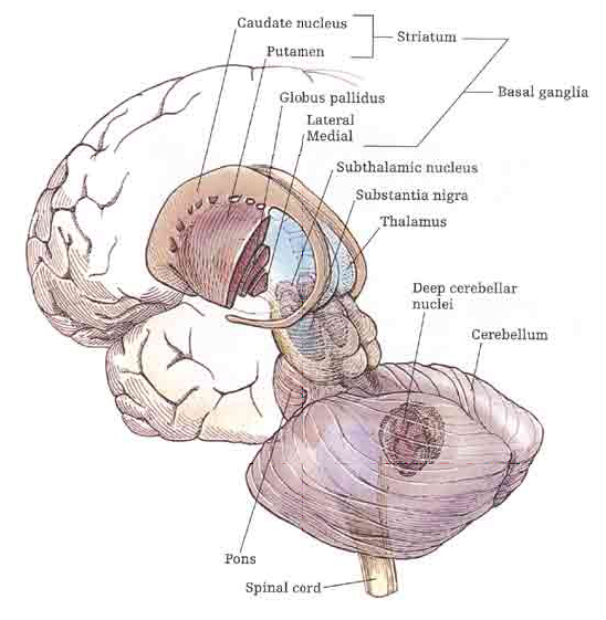
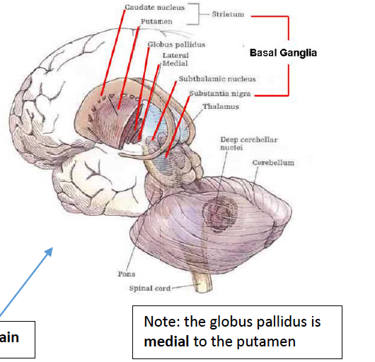
Basal Ganglia | Neupsy Key
Putamen And Globus Pallidus Form | Globus
Structure and Function of the Nervous System | Basicmedical Key
BrainMind.com
Caudate Putamen Globus Pallidus | Globus
Basal Ganglia | Basicmedical Key
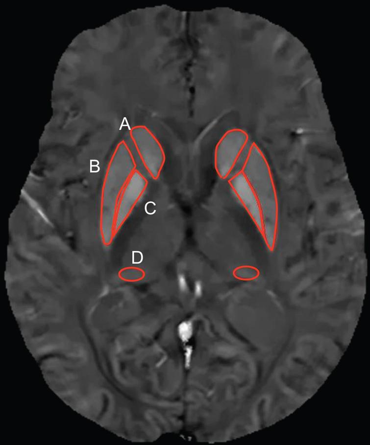
Representative SWI phase image with specific segmented structures …
Neuro content Flashcards | Quizlet
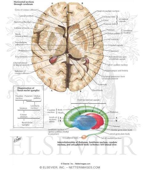
Basal Nuclei (Basal Ganglia) | GetBodySmart
Putamen And Globus Pallidus Mri | Globus
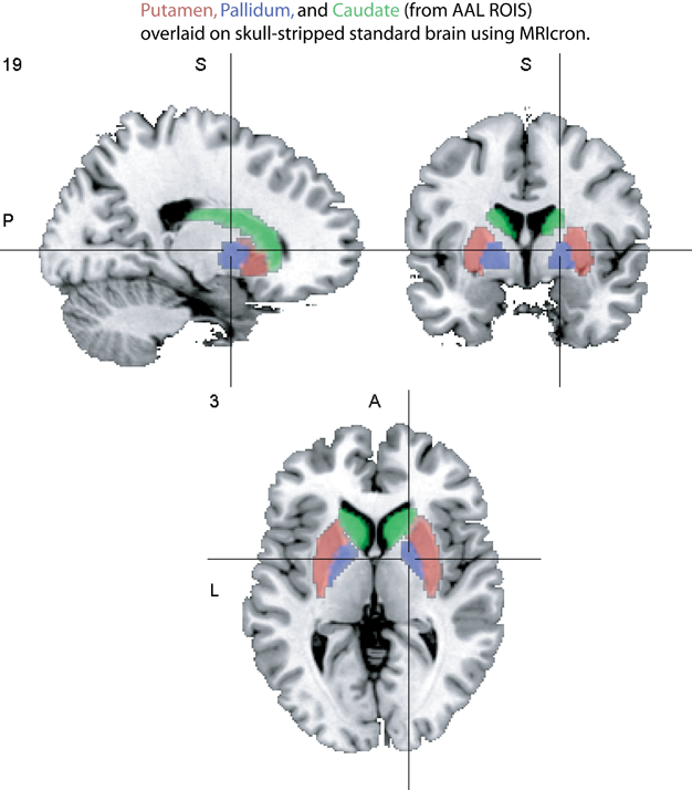
NeuroanatomyTutorial – MRC CBU Imaging Wiki
Globus Pallidus Putamen | Globus
Basal Ganglia, Striatum, Thalamus: Caudate, Putamen, Globus Pallidus …
Amygdaloid nucleus – grouped anatomically (sits on tail of caudate …
Putamen | Radiology Reference Article | Radiopaedia.org | Internal …
*caudate nucleus*- has a board head and slender tail. Tail extends …
Circulation of the Brain | Neupsy Key
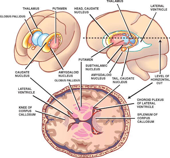
Illustration of the basal ganglia showing the caudate nucleus (red …
Basal ganglia – KINES 200: Introductory Neuroscience
Nucleus Caudate Functie
Brain Anatomy Globus Pallidus | Globus
coronal cut (important landmarks–thalamus, basal ganglia [caudate …
The basal ganglia are subcortical structures. The putamen and globus …
Caudate Putamen
Relationships of putamen, globus pallidus, and striatal iron …
Altered resting‐state functional connectivity of the putamen and …
T2 weighted FLAIR images of magnetic resonance scan showing bilaterally …
( A ) Subcortical ROIs in the right basal ganglia and thalamus. C …
KRS. Globus pallidus, caudate, and putamen hypointensity may be seen on …
A T1-weighted MRI image showing hyperintensity involving the right …
Localization of striatum (Caudate nucleus and putamen) in a rat brain …
Anatomy Of The Basal Ganglia, Neuroscience, Assignment Help
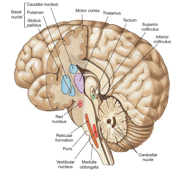
Basal Ganglia Basal Nuclei (Ganglia)
Globus Pallidus Putamen | Globus
Globus Pallidus Ct Head | Globus
(A-F) HO-1-positive glial cells within the caudate putamen (A-C …
Subthalamic Nucleus Deep Brain Stimulation: Basic Concepts and Novel …
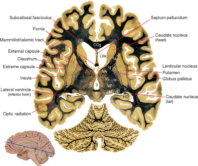
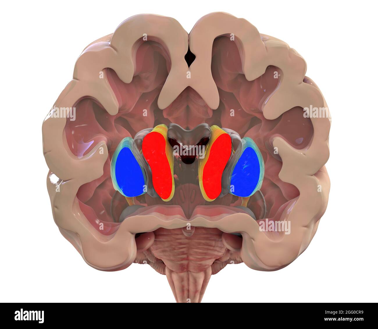
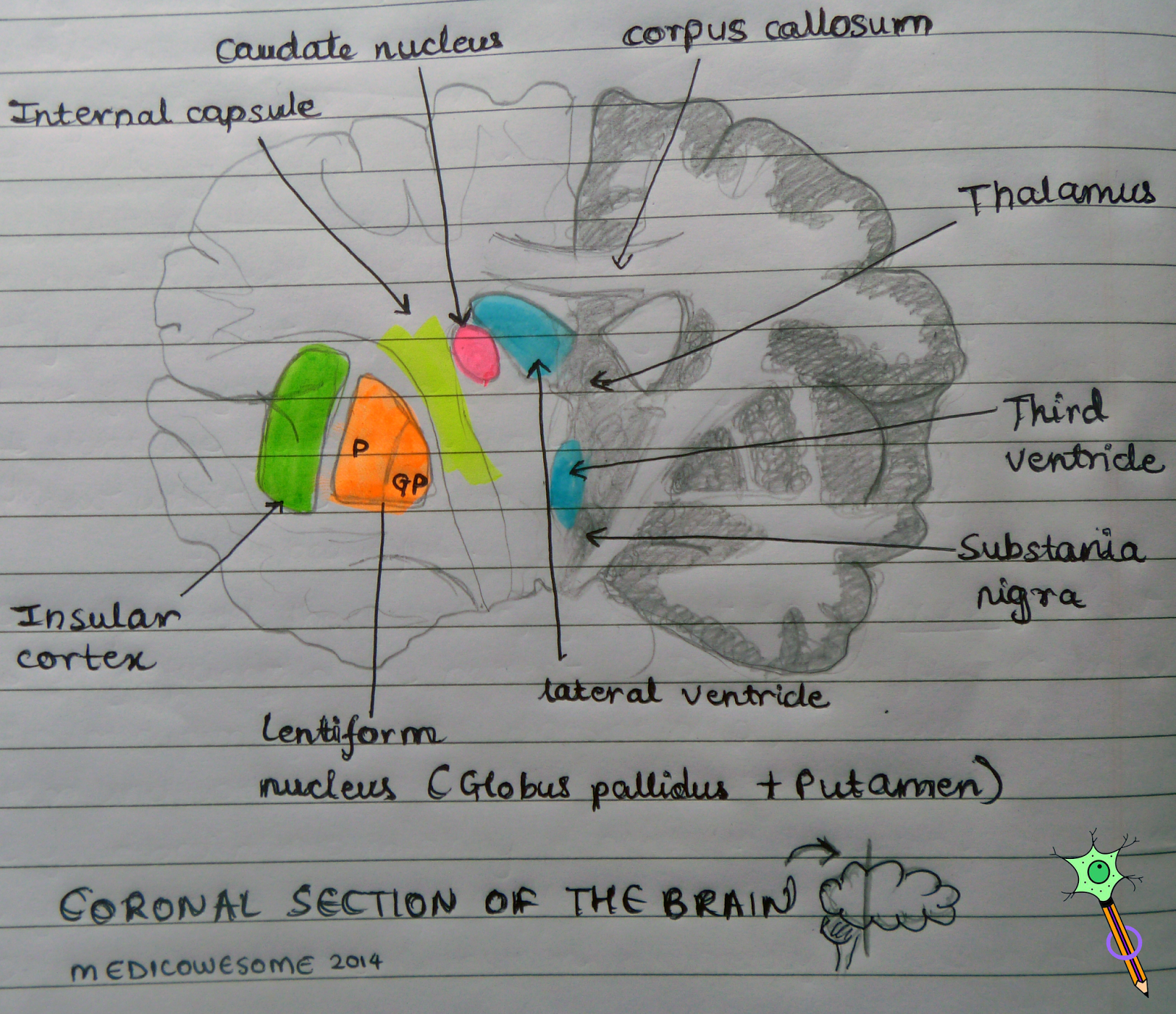
Medicowesome: Coronal section of the brain highlighting lentiform …
Example of exophytic iCM involving insular arteries and displacing …
harc 2 neuro Flashcards | Quizlet
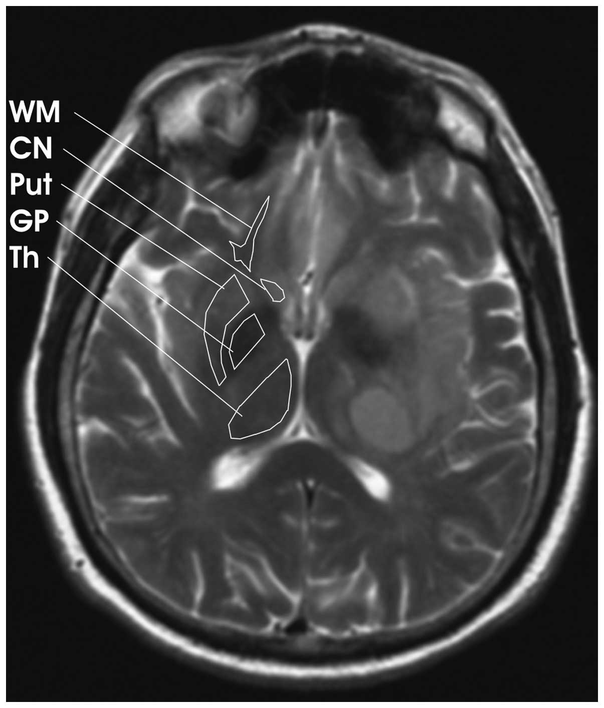
CN=Caudate nucleus, P=Putamen, GP=Globus pallidus, sn= substantia …
BrainMind.com
b. A PET scan, showing hypometabolism of the globus pallidus, caudate …
Basal Nuclei (Basal Ganglia) | GetBodySmart
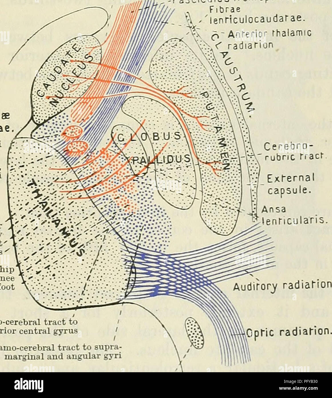
. Cunningham’s Text-book of anatomy. Anatomy. ,- Claustrum Insula …
Globus Pallidus Anatomy | Globus
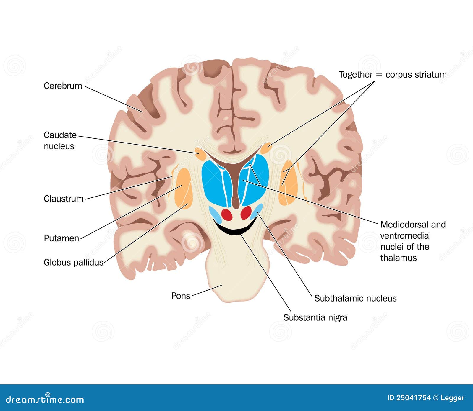
Anatomy Of Globus Pallidus In Basal Ganglia Royalty-Free Illustration …
basal ganglia Flashcards | Quizlet
Nucleus caudatus – Talot Suomessa
Putamen And Globus Pallidus Form | Globus
Studied regions of interest: caudate-putamen (CPu), hippocampus and …
9. Memory inhibition engaged caudate, putamen, and GPe more reliably …
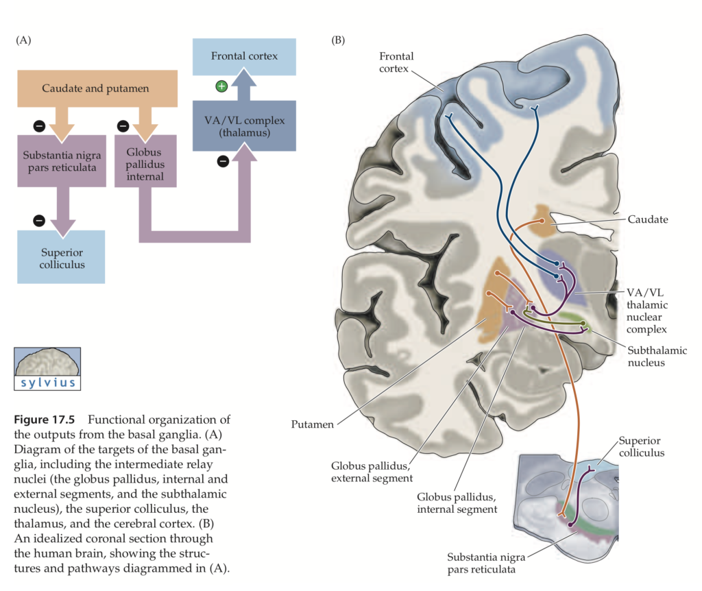
(a) Coronal section of a human brain showing the cortex, the caudate …
Anatomical connections of the PPN. SNr, substantia nigra; SNc …
CT scan without contrast which show caudate nucleous, pallidus and …
DAergic projections from the SNc and VTA innervate the caudate …
Basal Nuclei Circuit
FIRST segmentation. Example showing the seven subcortical regions …
Placements of ROIs for deep gray matters on T1-weighted image. 1 …
QSM images ( A and B ) and selected regions of interest ( C and D ). CA …
Calcifications of bilateral globus pallidus (black arrows). | Download …
(PDF) Prediction and classification of Alzheimer disease based on …
Striatal regions (bilateral dorsal putamen (z = 6 mm), right pallidum …
Reticular Formation and Limbic System – Textbook of Clinical …
Bidirectional arrows: pathways with both afferent and efferent …
A 3D rendering and axial MR images showing the subcortical grey matter …
Bilateral basal ganglia (caudate and putamen) hyperintensities on T2W1 …
腦梗塞(Cerebral Infarction) – 小小整理網站 Smallcollation
Brain Magnetic Resonance Imaging of Structural Abnormalities in Bipolar …
Figure 1:Histopathologic characterization of the BTBR mouse model of …
ROI analyses of right IFG, right putamen, right caudate, and right …
Neuroimaging of Diabetic Striatopathy: More Questions than Answers …
Basal Ganglia Flashcards | Quizlet
The Limbic System and Our Emotions | Limbic system, Brain structure …
Lecture 15 – Basal Ganglia Flashcards | Quizlet
Autoradiographs of rat brain at the level of caudate-putamen and …
Neuroanatomy Basal Ganglia (Nuclei) Diagram | Quizlet
Basal Ganglia | Neupsy Key
Medial Globus Pallidus Photograph by Sciepro/science Photo Library
2 Parallel loops among the basal ganglia, thalamus and cortex …
Basal Nuclei (Basal Ganglia) – Textbook of Clinical Neuroanatomy, 2 ed.
T2-weighted image showing bilateral symmetrical hyperintensities in the …
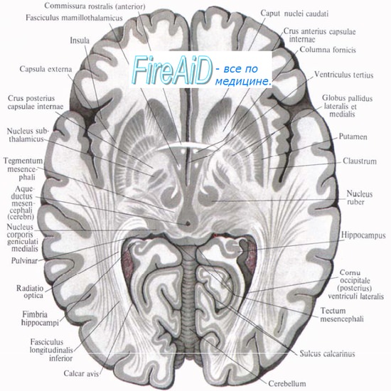
Анатомия: Стриопаллидарная система. Чечевицеобразное ядро, nucleus …
The macroscopic and microscopic findings in the patient. a, b The …
Principal fiber connections of the basal ganglia. Cx: Cerebral cortex …
Анатомия: Стриопаллидарная система. Чечевицеобразное ядро, nucleus …
Анатомия: Стриопаллидарная система. Чечевицеобразное ядро, nucleus …
Neuro – Basal Ganglia (Lecture 18) Flashcards | Quizlet
Pathology of the globus pallidus and dystonic postures of siblings with …
Patterns of Brain Iron Accumulation in Vascular Dementia and Alzheimer …
25 best Neuroanatomy images on Pinterest | Bing images, Neurology and …
Globus Pallidus Internus Deep Brain Stimulation for Disabling Diabetic …
Pallidum
Schematic diagram showing the glutamatergic pathways in the basal …
Figure 4 from Subthalamic Nucleus Deep Brain Stimulation: Basic …










:background_color(FFFFFF):format(jpeg)/images/library/5206/KOheX3BKhrTXAzS7z3TFDA_Globus_pallidus_01.png.jpeg)












:watermark(/images/watermark_only.png,0,0,0):watermark(/images/logo_url.png,-10,-10,0):format(jpeg)/images/anatomy_term/globus-pallidus-external-segment-1/DvTfHOpMcfK90bumzWE3Q_20Globus_pallidus_external_segment.png)


