List showcases captivating images of which condition is a swelling and hyperemia of the optic disc, also called choked disc? gathered and meticulously curated by the website galleryz.online. Furthermore, you can find more related images in the details below.
which condition is a swelling and hyperemia of the optic disc, also called choked disc?
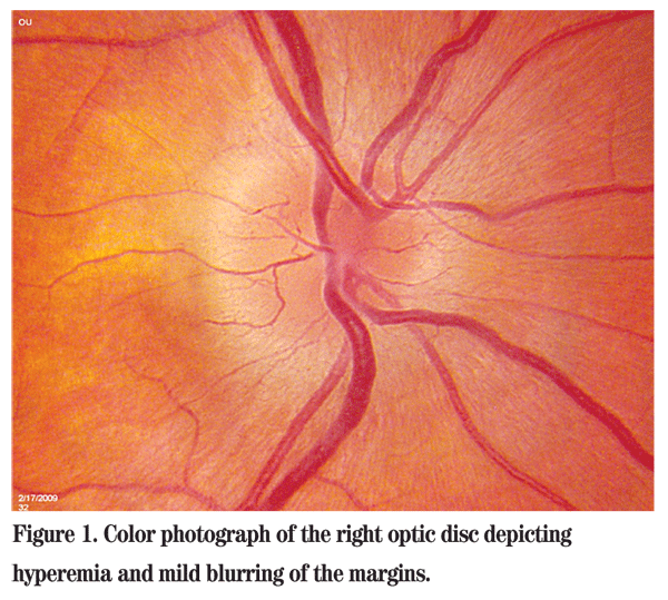
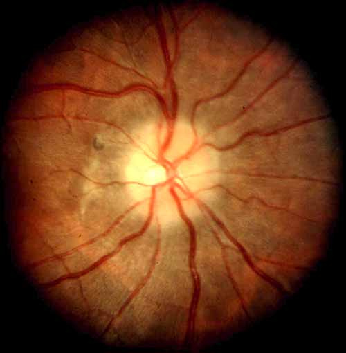
 Right fundus photograph showing optic disc edema and hyperemia, with a …
 Blurred margins, hyperemia, and swelling of the optic disc | Download …
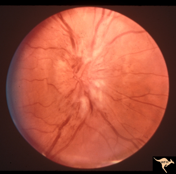
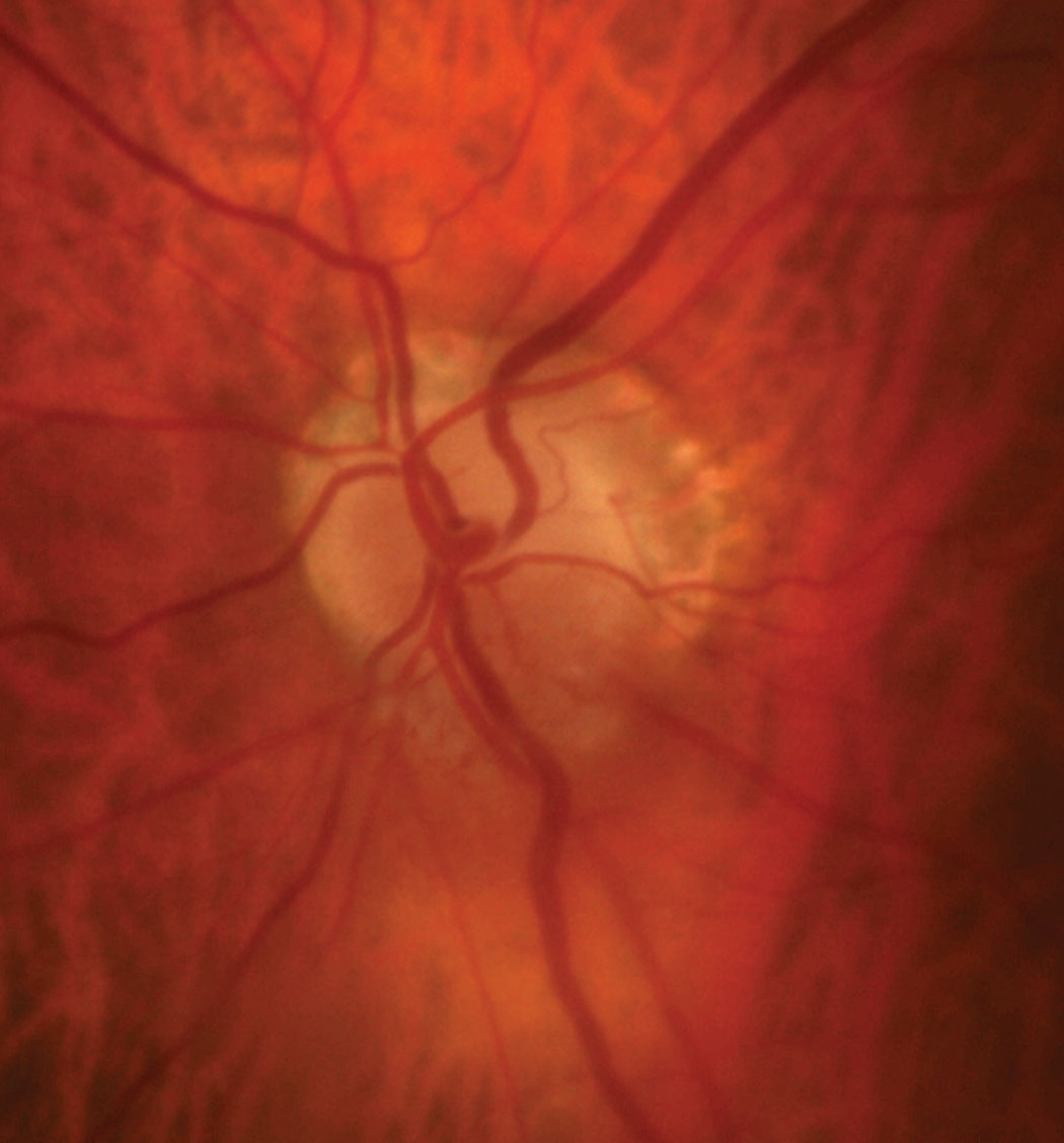 What’s Your Disc Diagnosis?
1,366 – Chocolate Chip Ear Wax Disc Removal
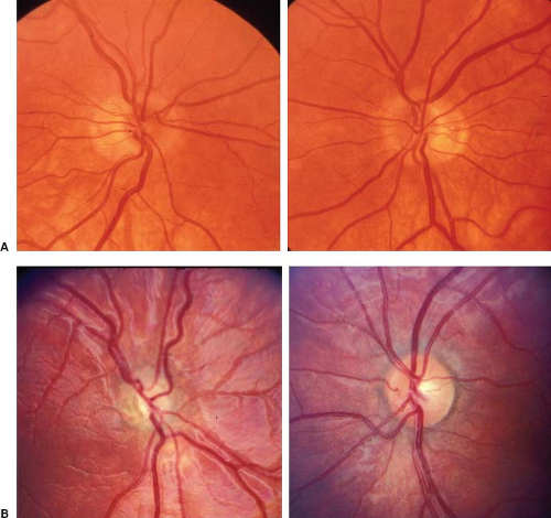
 (A) Colour photograph of right posterior pole (case 1, 20 year old man …
 (Case 4). Pale disc swelling of the optic disc is noted in the right …
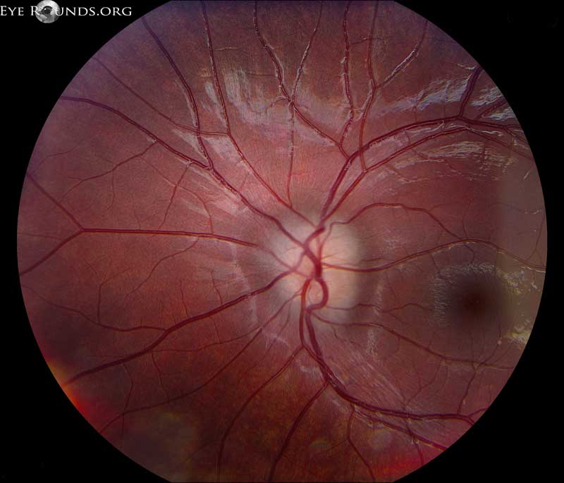
 Left fundus photograph showing optic disc edema and hyperemia during …
 Figure1. a) Appearance of the right optic disc in non-arteritic …
 Pin on Eye: Dots, Spots & lines
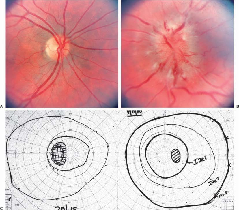
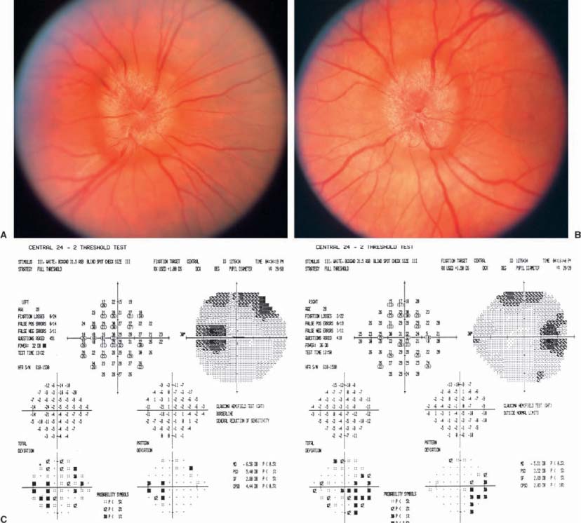
 Fundus abnormalities at presentation. (a) Swelling optic disc with …
 Papilloedema: an update | Eye News
 Fundus picture of both eyes showed optic disc edema in the right eye …
 (A) Fundus photograph of the right eye shows swelling of the disc and …
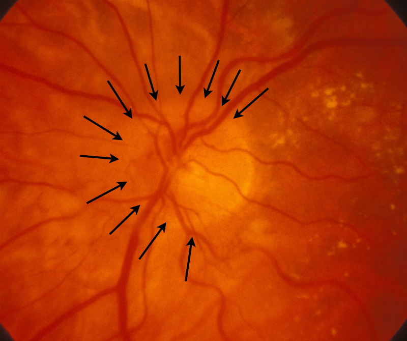
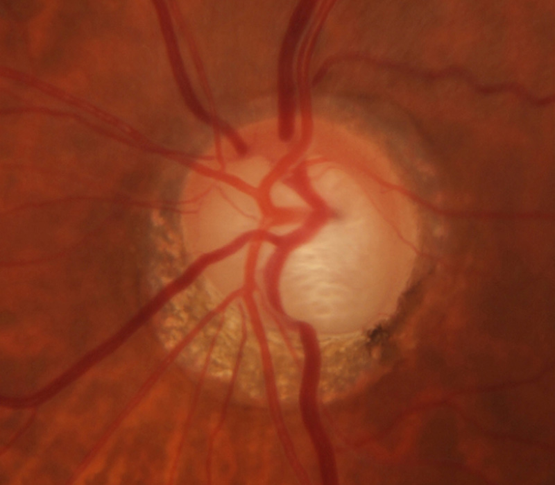
 Right eye color fundus picture at onset (a) Optic disc hyperemia and …
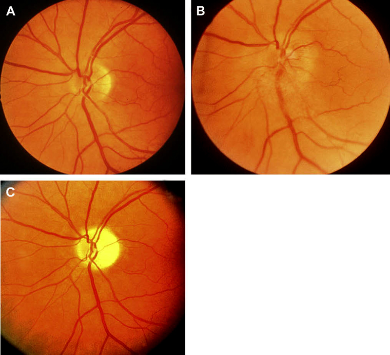
 Examples of optic disc swelling by the Frisén grading scheme. a Frisén …
 Swollen optic disk – American Academy of Ophthalmology
 High Incidence of Optic Disc Swelling at Very High Altitudes | Neuro …
 Optic disc photographs of a 49-year-old male with nutritional optic …
 Normal appearing optic disc is seen in all cases of acute posterior …
 Optic disk edema in cyclosporine (CsA) toxicity. Reprinted with …
 Bilateral Optic Disc Swelling: Presenting Sign of Pheochromocytoma in Child
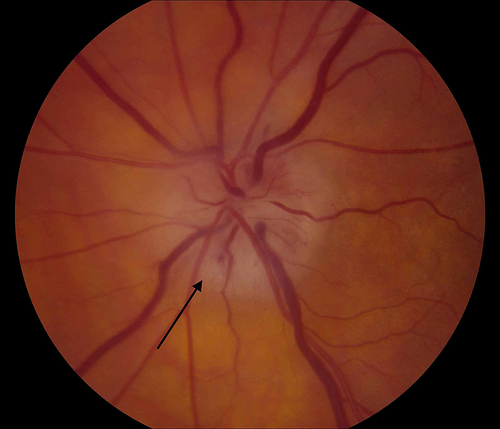
 Fundus photograph showing disc hyperaemia with attenuation of retinal …
 Postflight fundus examination photos (bottom) show grade 3 and grade 1 …
 Swollen optic disc of the right eye with small hemorrhages. | Download …
 Pin on Медицина
 Case 2. Fundus photograph of the left eye, showing diff use pale optic …
 F2b05 Optic Disc Swelling from Optic Glioma | Eccles Health Sciences …
 Optic disc hemorrhages in glaucoma and common clinical features …
 (A) The optic nerve head is swollen with blurring of the disc margin …
 Differential diagnosis of bilateral optic disc swelling | Download Table
 Wills Resident Case Series
 A. Both right (R) and left (L) optic discs show acute edema, hyperemia …
 Case 2 right optic disc photograph, 10 days after onset of visual loss …
 Case 2 right optic disc photograph, 10 days after onset of visual loss …
 Anterior Ischemic Optic Neuropathy: part 2. A discussion for physicians
 Optic disc edema. COMS Grading
 Optic Neuritis | Optic neuritis, Optometry education, Opthalmic technician
 The Swollen Optic Disc in Children | SpringerLink
 Fundus photograph showing the optic disc—fovea line (black line), outer …
 Moran CORE | Disc Edema
 medical images M-P | medicalimages
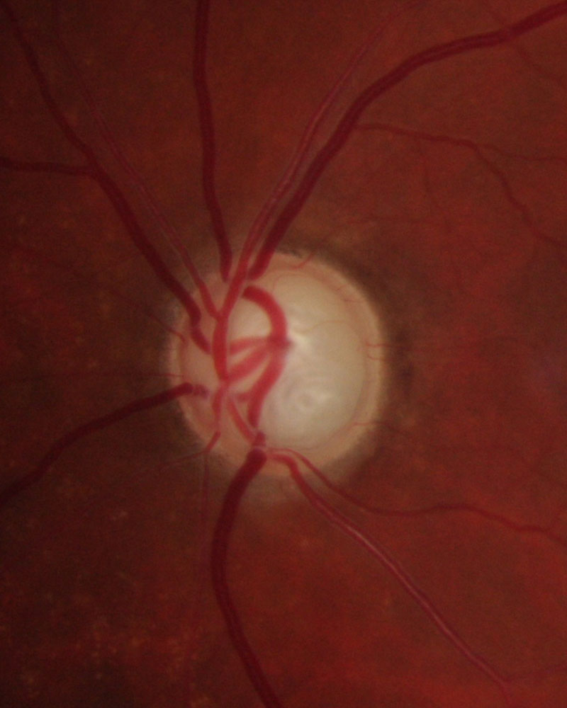
 Optic disc pit OCT image – American Academy of Ophthalmology
 Optic disc showing disc hemorrhage (Rim to disc ratio | Download …
 Glaucoma Information Glaucomatous optic disc – Glaucoma Information
 CMDT Media Library | AccessMedicine | McGraw-Hill Medical
 | (a) Right eye with normal appearance of the optic nerve and macula …
 Fundus imaging. FFA and OCT showed the swollen optic disc and optic …
 Lecture: Approach to the Patient with Bilateral Optic Disc Swelling …
 Pseudotumor Cerebri; Benign Intracranial Hypertension; Idiopathic …
 Optic disc appearance, case 2. Fundoscopy of the left eye (a) and right …
 optic disc fovea – ค้นหาด้วย Google | OSCE | Pinterest | Search
 Cerebral small vessel disease in a patient with NAION. (A) A case of …
 Optic Disc Hemorrhage – EyeWiki
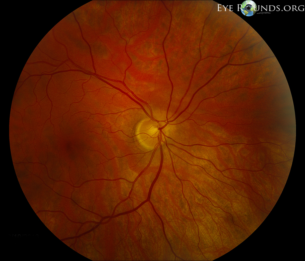
 Presence of hyperaemic and swollen optic disc with pre-retinal …
 Retinography showing mild hyperemia of both optic discs. | Download …
 Optic atrophy | Ento Key
 Optic Disc Edema | Ento Key
 Optic Nerve Diseases in Children – Eye Specialist, Treatments
 Fundus photograph of the right eye of a low-flow CCF patient showing …
 Optic Disc Edema and Hemorrhages with Subdural Hematoma – Retina Image Bank
 Comparisons of the fundus photograph and OCT pairs with mild optic disc …
 Optic discs appearance and optical coherence tomography (OCT) findings …
 Moran CORE | A Case Report of Hypertensive Retinopathy
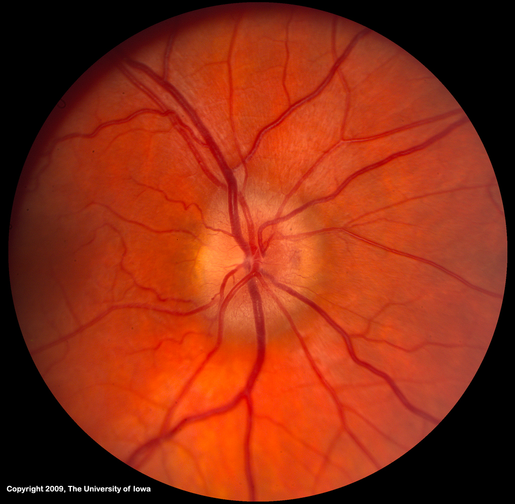
 Normal Optic Disc – Physical Diagnosis – Mitch Medical Healthcare
 Macula, Optic Disc and Blood Tree in a Fundus Photography. Image by …
 Left eye, 9 months later. a Color fundus picture showing the quietish …
 The swollen optic nerve: an approach to diagnosis and management …
 Causes of Optic Nerve Edema – Los Alamos Family Eyecare, P.C.
 a Fundus photograph showing diffuse optic disc pallor in both eyes …
 Services — Lone Star Eye
 Congenital Anomalies of the Optic Disc | Ento Key
 Ischemic optic neuropathy (ION) | MedLink Neurology
 Normal Tension Glaucoma – EyeWiki
 Optic disc edema. COMS Grading
 Optic Disc Edema | Ento Key
 Fundus imaging showed (a) a swollen optic disc in the right eye, (b) a …
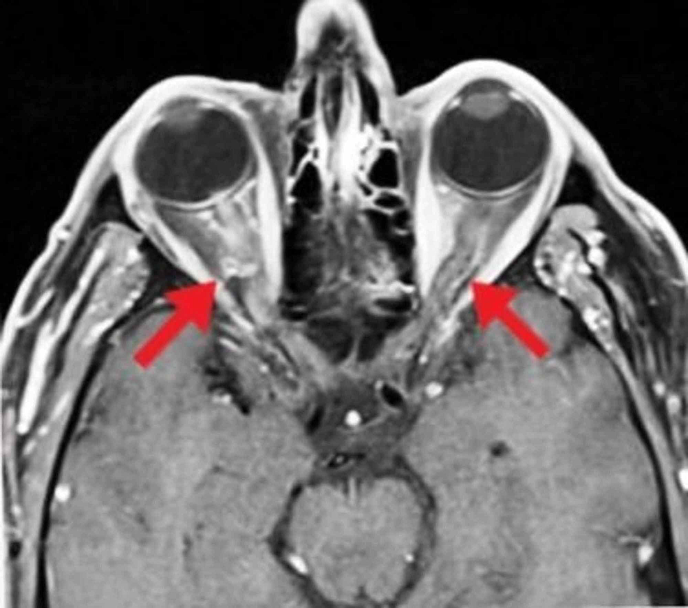
 Cureus | High-Sensitivity C-Reactive Protein and Magnetic Resonance …
 Congenital Optic Disc Anomalies — Ophthalmology Review
 Examples of Left versus Right and Macular versus Optic Disc-centred …
 Tilted Disc Syndrome – EyeWiki
 Community Eye Health Journal » Defining and diagnosing glaucoma: a …
 Typical fundal appearance in dominant optic atrophy showing bilateral …
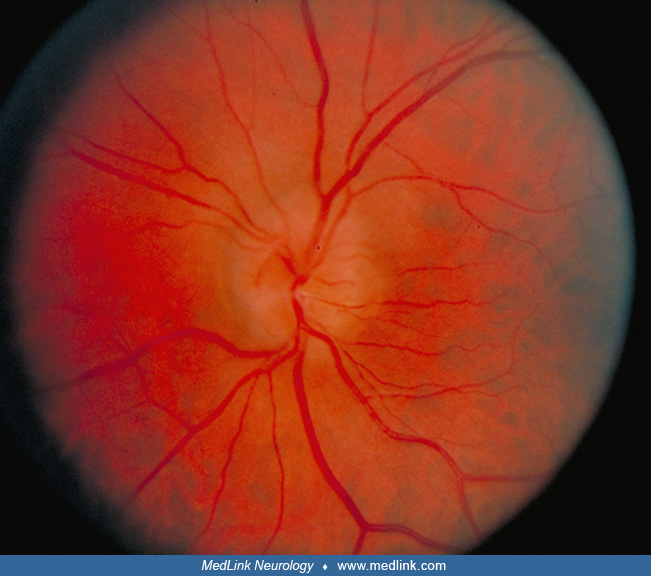 Ischemic optic neuropathy (ION) | MedLink Neurology
 (A) Ophthalmoscopic findings at the time of diagnosis of ARN. Optic …
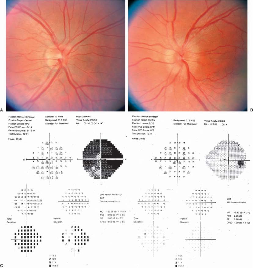
 Differentiation between optic disc drusen and optic disc oedema using …
 Causes Of Enlarged Blind Spot – BLINDS
 The Swollen Optic Disc in Children | SpringerLink
 Congenital Optic Disc Anomalies | SpringerLink
 Cureus | Bilateral Optic Neuritis: A Rare Complication of Mumps
 Idiopathic Intracranial Hypertension: pseudotumor cerebri
 Myopic Shift With Tilted Optic Disc. – Retina Image Bank
 An International Treatment Trial for Patients with NAION
 Clinical Reasoning: An unusual pattern of optic disc swelling and …
 Disc swelling. Diabetic papillopathy | Eccles Health Sciences Library …
 Atlas Entry – Anomalous appearing optic nerves in both eyes